Circulating melanoma exosomes as diagnostic and prognosis biomarkers
Circulating melanoma exosomes as diagnostic and prognosis biomarkers
Estibaliz Alegre a ,Leyre Zubiri b ,Jose Luis Perez-Gracia b ,María González-Cao c ,Lourdes Soria b ,Salvador Martín-Algarra b ,Alvaro González a ,?
a Laboratory of Biochemistry,University Clinic of Navarra,Spain
b Department of Medical Oncology,University Clini
c of Navarra,Spain
c
Translational Cancer Research Unit,Instituto Oncológico Dr Rosell,Quirón Dexeus University Hospital,Barcelona,Spain
a b s t r a c t
a r t i c l e i n f o Article history:
Received 16November 2015
Received in revised form 11December 2015Accepted 21December 2015
Available online 24December 2015Background:Malignant melanoma is an aggressive cancer with an increasing incidence.Exosomes are actively se-creted microvesicles,whose characteristics re ?ect those of the cell they are originated in.The aim of this study was to identify and evaluate the presence of the melanoma biomarkers MIA,S100B and tyrosinase-related pro-tein 2(TYRP2)in exosomes and their potential clinical utility.
Methods:Serum samples were obtained from stage IV melanoma patients,melanoma-free patients and healthy controls.Exosomes were precipitated and TYRP2,MIA and S100B concentrations were quanti ?ed in serum,exosomes,and exosome-free serum.
Results:Both MIA and S100B were detected in exosomes and correlated signi ?cantly with serum concentrations (S100B:r =0.968;MIA:r =0.799;p b 0.001).MIA and S100B concentrations in exosomes were signi ?cantly higher in melanoma patients than in healthy controls and disease-free patients.However,TYRP2concentrations in exosomes did not differ between these three groups.ROC curves analysis rendered AUCs for MIA of 0.883(p b 0.01)and of 0.840for S100B (p b 0.01).Patients with exosome MIA concentration higher than 2.5μg/L showed shorter median survival related to those with lower level (4versus 11months;p b 0.05).
Conclusions:MIA and S100B can be detected in exosomes from melanoma patients and their quanti ?cation pre-sents diagnostic and prognostic utility.
?2015Elsevier B.V.All rights reserved.
Keywords:
Melanoma inhibitory activity S100B Exosomes Melanoma
1.Introduction
Metastatic melanoma is a very aggressive cancer whose incidence is increasing worldwide.The prognosis is generally poor,although new treatments have improved recently overall survival.S100B,Melanoma Inhibitory Activity (MIA)and Lactate Dehydrogenase (LDH)are the most widely used tumor markers for prognosis and follow-up in ad-vanced melanoma [1,2].MIA is a small soluble protein of 11kDa secret-ed by malignant melanoma cells.S100is a 21kDa dimeric protein composed of 2subunits,αor β,being the αβheterodimer expressed by melanoma cells [3].LDH concentrations higher than reference range can classify patients with metastatic melanoma in a more ad-vanced stage (M1c)[4].Both MIA and S100B serum concentrations are elevated in advanced melanoma and their measurement can be use-ful as prognostic factors and to monitor the disease in stages III and IV
[1,5].However they show some limitations of speci ?city and sensitivity,especially LDH and therefore,they are not widely used [2,6,7].
Recently,other biomarkers have been investigated,such as cell-free nucleic acids and exosomes.BRAF mutations in cell-free DNA have also shown to be useful to monitor melanoma patients treated with BRAF in-hibitors [8,9].Exosomes are actively secreted microvesicles derived from the cellular endosomal membrane with sizes ranging from 30to 200nm [10].Cancer cells,and particularly melanoma cells,can release large quantities of exosomes [11],in contrast to normal melanocytes [12].These exosomes derived from cancer cells participate in tumor progression with immune-suppressive functions [13],contributing to angiogenesis [14],drug resistance [15]and cell migration [16].As a re-sult,exosomes are now considered to play a pivotal role in tumor devel-opment and progression [17].
The composition of nucleic acids and proteins cargo of exosomes re-?ects the cells they originate from [18].In fact,Peinado et al.[17]found that exosomes isolated from stage IV melanoma patients characteristi-cally contain the proteins tyrosinase-related protein 2(dopachrome tautomerase,TYRP2),Very Late Antigen 4,Heat Shock Protein 70,HSP90isoform,and MET oncoprotein [17].In addition,melanoma exosomes seem to play a role in the metastasis to lymph nodes.Partic-ularly,TYRP2expression in exosomes has been associated with meta-static progression in advanced melanoma [17].
Clinica Chimica Acta 454(2016)28–32
Abbreviations:AUC,Area Under Curve;LDH,Lactate Dehydrogenase;MIA,Melanoma Inhibitory Activity;ROC,Receiver Operating Characteristics;TYRP2,tyrosinase-related protein 2,Dopachrome tautomerase.
?Corresponding author at:Laboratory of Biochemistry,University Clinic of Navarra,Av.Pio XII 36,Pamplona 31008,Spain.
E-mail address:agonzaleh@unav.es (A.
González).
https://www.360docs.net/doc/9214082795.html,/10.1016/https://www.360docs.net/doc/9214082795.html,a.2015.12.031
0009-8981/?2015Elsevier B.V.All rights
reserved.
Contents lists available at ScienceDirect
Clinica Chimica Acta
j o u rn a l h o m e p a g e :w w w.e l s e vi e r.c o m/l o c a t e /c l i n c h i m
Tumor-derived exosomes have been detected in several biological ?uids and can carry tumor speci?c antigens,such as carcinoembryogenic antigen in ascitic exosomes from colon carcinoma[19],prostate speci?c antigen in urinary exosomes from prostate cancer[20],or CA125in ascitic exosomes from ovarian carcinoma[21].The expression of tumor-speci?c antigens by exosomes can render these particles useful for cancer diagno-sis and monitoring.For this reason,the aim of the present work was to study the presence of melanoma biomarkers S100B and MIA in exosomes and to compare its utility with their determination in serum.
2.Material&methods
2.1.Sample collection
Peripheral blood samples were collected after obtaining informed consent from53advanced melanoma patients(mean age:55years; males:54%),18melanoma disease-free patients(mean age:54years; males:43%)and from25healthy volunteers(mean age:41years; males:29%)used as age and sex matched control groups.Blood samples were centrifuged and serum was isolated and kept at?80°C until anal-ysis.The study was approved by our institution's Ethical Review Board.
M8and UMBY melanoma cell lines were cultured in RPMI1640me-dium supplemented with2mmol/L L-glutamine,10%exosome-free heat inactivated fetal bovine serum,penicillin G100U/mL,geniticin 1mg/mL,streptomycin100μg/mL(Gibco,Grand Island,NY,USA),and were cultured in a37°C,5%CO2humidi?ed incubator.
2.2.Exosome isolation
Exosomes were obtained from serum with ExoQuick precipitation solution(System Biosciences,Mountain View,CA,USA)according to manufacturer's instructions.Brie?y,250μL,of serum were mixed with 63μL of ExoQuick?solution and incubated for1h at4°C.After centri-fugation at1500g for30min,pellets were suspended in100–200μL of PBS or in lysis buffer.
Exosomes from cell cultures were collected by ultracentrifugation at 110,000g for2h at4°C after precleaning the media by two successive centrifugations and?ltration through a0.22μm?lter(EMD Millipore, Billerica,MA,USA).Pellets were resuspended in PBS or in lysis buffer.
2.3.Exosome size analysis
Isolated exosomes were diluted in PBS and particle size was ana-lyzed using Malvern Zetasizer Nano-ZS(Malvern Instruments,UK) and its corresponding software(Zetasizer Ver.7.03).
2.4.Protein concentration
Protein concentration in exosomes was estimated through absor-bance at280nm with a Nanodrop spectrophotometer(Thermo Fisher Scienti?c,Wilmington,USA).A correction factor was introduced to cor-rect sample volume reduction from250to200μL.
2.5.Western blot
Proteins(20μg)were denatured at100°C for5min in loading buffer containing125mM Tris(pH 6.8),4%SDS,30%glycerol,5%β-mercaptoethanol and0.4%bromophenol.Proteins were subjected to 10%polyacrylamide gel electrophoresis under denaturing conditions (SDS-PAGE),with subsequent electroblotting transfer onto a nitrocellu-lose membrane.The membrane was blocked with5%skimmed milk in PBS-Tween0.1%during30min at room temperature,and then incubat-ed during1h with anti-CD63antibody(Santa Cruz Biotechnology, Texas,USA)diluted1:1000in PBS-Tween0.1%.Immunoblot analysis was performed using an HRP-conjugated anti-mouse antibody (1:5000;Amersham Biosciences,Uppsala,Sweden),and developed using the ECL kit(Amersham Biosciences).
2.6.Immuno assays
MIA was determined by a manual commercial enzyme-linked im-munosorbent assay(Roche,Mannheim,Germany)and S100B was ana-lyzed by an electrochemiluminescence assay automatized in an e602 Module from a C8000(Roche Diagnostics,Mannheim,Germany)[1]. Tyrosinase-related protein2concentration was measured using a com-mercial ELISA kit(Cloud-Clone Corp,Houston,USA).This is a sandwich enzyme immunoassay that uses two antibodies speci?c for TYRP2.A correction factor was introduced to correct sample volume reduction.
The concentrations of biomarker in exosome-free serum were calcu-lated by subtracting the measured concentration in exosomes to the serum concentration.
2.7.Statistical analysis
Concentrations were expressed as median and25th–75th percentile after determining their non-Gaussian distribution with the Kolmogo-rov–Smirnov and Shapiro–Wilk's tests.The non-parametric Kruskal–Wallis test and Dunn's multiple comparison test were applied to com-pare the levels of the tumor markers.Correlation analysis was per-formed using the Pearson correlation test.Overall survival was measured from the time of blood drawing to death or last follow-up and it was analyzed by the Kaplan–Meier method and compared by the Gehan–Breslow–Wicoxon test.Receiver operating characteristic (ROC)curves were constructed to determine the sensitivity and speci-?city of the assays.A two-tailed p-value b0.05was considered to be sta-tistically signi?cant.Statistical analysis was performed with GraphPad Prism version6.07(La Jolla,CA,USA).
3.Results
3.1.Identi?cation of serum exosomes
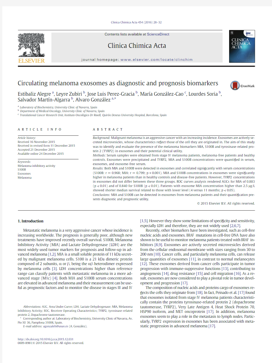
The mean size of the microvesicles obtained by precipitation with ExoQuick and by ultracentrifugation was lower than200nm,similar to that described for exosomes[10](Fig.1A).Exosomes isolated from plasma were characterized by Western blot using anti-CD63and,as ex-pected,we observed the predicted band at53kDa(Fig.1B).In conclu-sion,we could identify these microvesicles obtained by precipitation as exosomes.
The concentration of exosomes isolated using the ExoQuick reagent was estimated by measuring the protein concentration.The median concentration of proteins in exosomes from melanoma patients was 8μg/L(Q1–Q3:6–10μg/L),very similar to the control group(median: 9μg/L;Q1–Q3:8–10μg/L).These results indicate that patients with ad-vanced melanoma do not show signi?cant differences in serum exosome levels,as compared to melanoma-free patients and healthy controls.
3.2.Biomarkers analysis in exosomes from melanoma patients
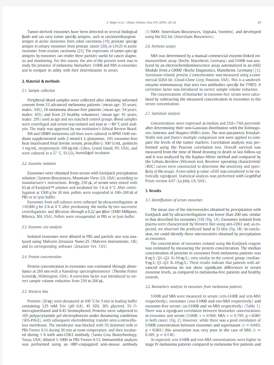
S100B and MIA were measured in serum(srm-S100B and srm-MIA respectively),exosomes(exo-S100B and exo-MIA respectively)and exosome-free serum(sn-S100B and sn-MIA respectively)(Table1). There was a signi?cant correlation between biomarker concentrations in exosomes and serum(S100B:r=0.968;MIA:r=0.799;p b0.001 in both cases)(Fig.2).However,while there was a good correlation of S100B concentration between exosomes and supernatant(r=0.943; p b0.001)this association was very poor in the case of MIA(r= 0.189;p=0.138).
As expected,srm-S100B and srm-MIA concentrations were higher in stage IV melanoma patients compared to melanoma free patients and
29
E.Alegre et al./Clinica Chimica Acta454(2016)28–32
healthy controls.S100B and MIA levels in exosomes were also statisti-cally higher in advanced melanoma patients,as compared to both healthy controls and melanoma free-patients (p b 0.01in both cases,Fig.3,Table 1).
To know if there was a change in the secretion of these biomarkers in exosomes,we studied the percentage of S100B or MIA concentrations in exosomes in relation to their serum levels.We observed a signi ?cant decrease in percentage of S100B in exosome from patients in stage IV (median stage IV:17%,Q1–Q3:12%–21%)compared to controls and dis-ease free patients (median control:23%,Q1–Q3:20%–26%;median dis-ease free:24%,Q1–Q3:18%–29%;p b 0.05related to stage IV)(Fig.3).This difference was not observed when MIA was analyzed (median con-trol:18%,Q1–Q3:6%–23%;median disease free:17%,Q1–Q3:14%–26%;median stage IV:17%,Q1–Q3:12%–28%).
Finally,we analyzed the potential utility of TYRP2concentrations in exosomes.There was a signi ?cant correlation in its levels between serum and exosomes (r =0.941;p b 0.01).The percentage of TYRP2in exosomes (median:61%;Q1–Q3:54%–68%;p b 0.01)was signi ?cant-ly higher than the percentage of MIA in exosomes (median:17%;Q1–Q3:11%–26%)or the percentage of S100B in exosomes (median:21%;
Q1–Q3:14%–25%).However,contrary to what occurs with S100B and MIA,we did not detect differences in either serum or exosome levels be-tween control and melanoma patients (Table 1).
These results show that both S100B and MIA concentrations in exosomes from melanoma patients are higher than in healthy controls and disease free patients.
3.3.Diagnostic and prognostic value of S100B and MIA concentrations in exosomes
Next,we analyzed the sensitivity and speci ?city of exo-MIA and exo-S100B to discriminate advanced melanoma patients from healthy con-trols.The ROC analysis demonstrated an AUC of 0.883(95%CI =0.808–0.959;p b 0.01)for exo-MIA,better than srm-MIA (AUC:0.806;95%CI =0.701–0.903;p b 0.01)(Fig.4).With a cut-off value of exo-MIA set at 1.4μg/L,the sensitivity and the speci ?city was 80%.Also,the ROC for exo-S100B had an AUC of 0.840(95%CI =0.745to 0.936;p b 0.01),better than that observed for srm-S100B (AUC:0.806;95%CI =0.709to 0.902;p b 0.01).With a cutoff value of exo-S100B set at 0.015μg/L,the sensitivity and speci ?city was 80%.
Concentrations of MIA in exosomes above the median value were as-sociated with shorter survival (Fig.5).Patients with exo-MIA levels equal or higher than 2.5μg/L showed a median survival of 4months,while patients with lower concentrations presented a median survival of 11months (p b 0.05).Also,patients with exo-S100B concentrations higher than 0.03μg/L showed a median survival of 7months,while pa-tients with lower levels had a median survival of 10months (p =0.296).4.Discussion
We have shown that both S100B and MIA are detected in exosomes obtained from melanoma patients.The method used here to measure S100B employs monoclonal antibodies directed against the β-chain,so it can detect both the AB-and BB-dimers [22].Other proteins of the S100family have been identi ?ed in most proteomic analysis
of
Fig.1.A:Representative experiment (n =4)of size distribution of exosomes obtained from serum and from M8melanoma cell line.B:Representative experiment (n =5)of Western blot incubated with anti-CD63of serum exosomes and of the whole serum.
Table 1
Serum (srm-),exosome (exo-)and exosome-free serum (sn-)levels of MIA,S100B and TYRP2in healthy controls,melanoma-free patient s,and stage IV melanoma patients.Data are expressed as median and interquartile range.P b 0.05.
Controls
Disease free Stage IV
srm-MIA (μg/L)6(5–7) 5.2(3.9–6.6)14(7–45)a ,b exo-MIA (μg/L)1(0.4–1.5)1(0.9–1.7) 2.5(1.5–7.2)a ,b sn-MIA (μg/L) 4.9(4–6)
4.1(3–5)
10(5–33)a ,b
srm-S100B (μg/L)0.06(0.05–0.07)0.04(0.03–0.07)0.22(0.07–0.67)a ,b exo-S100B (μg/L)0.01(0.01–0.02)0.01(0.0–0.02)0.04(0.02–0.09)a ,b sn-S100B (μg/L)0.04(0.04–0.05)0.03(0.02–0.06)0.23(0.06–0.60)a ,b srm-TYRP2(μg/L)13(11–16)12(6–22)9(8–22)exo-TYRP2(μg/L)8(7–9)9(5–11)6(5–11)sn-TYRP2(μg/L)
5(4–7)
3(1–10)
3(2–11)
a Related to control.
b
Related to disease free
patients.
Fig.2.Relationship between MIA (A)and S100B (B)concentrations in exosomes (exo-)and either serum (srm-)or exosome-free supernatant (sn-).R2coef ?cient is indicated for each relationship and p b 0.05was considered signi ?cant.
30 E.Alegre et al./Clinica Chimica Acta 454(2016)28–32
exosomes.S100A1,S100A6and S100A11,which can interact with S100B forming heterodimers [23],have been particularly detected [24].Furthermore,we could also detect the melanoma biomarker S100B by ELISA in exosomes from M8and UMBY melanoma cell lines (data not shown).To our knowledge,this is the ?rst report in which S100B and MIA have been documented in exosomes.However,the pro-portion of S100B in exosomes decreased in stage IV patients which can be due to S100B released by melanoma cells mainly by necrosis and ap-optosis and not due to overexpression or active secretion [25].This was not observed in the case of MIA.MIA is a secreted protein that binds to integrins,particularly α4β1and α5β1[26].These integrins and others are present in exosomes and MIA could be bound to these proteins.Both S100B and MIA,which are well known biomarkers in melanoma [1],can be used to identify exosomes released by melanoma cells.
We have observed that the sensitivity and speci ?city for melanoma of the S100B or MIA assays in exosomes are superior to their correspon-dent analysis in serum.By measuring S100B and MIA in exosomes,in-terferences from other sources can be avoided.With this approach we were able to detect only those proteins selectively delivered,and we ex-cluded those proteins released by cell death,as they are not included in exosomes.Also,apoptotic bodies,debris and other microvesicles that could contain these proteins are usually in the range of 1μm [27],so
they are eliminated in the process of exosome isolation.For this reason,the quanti ?cation of these molecules in exosomes could increase the speci ?city of the biomarker,at the same time that it could permit to identify those exosomes derived from the tumor.Other authors have shown that the analysis of speci ?c tumor proteins in exosomes could improve the sensitivity and speci ?city to detect cancer patients.Peng et al.detected CA125located on the surface of exosomes,which allowed one to identify the exosomes from tumor cells in the ascites of ovarian cancer patients [21].Also,recently,it was shown that Glypican-1in cir-culating serum exosomes can differentiate patients with pancreatic can-cer from healthy subjects or patients with a benign pancreatic disease with absolute speci ?city and sensitivity [28].However,further studies in benign conditions other than melanoma where MIA [29]or S100B [30]are elevated,should be performed [2].As we and others have pre-viously shown in case of serum [1,5,31],it seems that not only does MIA present improved sensitivity and speci ?city in melanoma patients,but also has prognostic value,since advanced stage patients with exo-MIA concentrations higher than 2.5μg/L presented shorter overall survival than patients with lower levels.
Previous authors have shown increased concentrations of the house-keeping exosome proteins CD63and Rab-5b in melanoma patients as compared with healthy controls [32].We have not detected differences in exosome levels between melanoma patients and healthy controls.Probably these discordances could be explained by the different methods used to obtain and measure exosomes.Most other studies an-alyzing proteomic pro ?le in exosomes employ cumbersome technology,such as ultracentrifugation and mass-spectrometry [12,16,17].Impor-tantly,we have performed this study using not very time-consuming technologies available in most clinical laboratories so our methodology can be easily translated into routine [33].Exosomes from clinical sam-ples can be easily obtained using ExoQuick method [18],a technology that captures these microvesicles in “polymer nets ”[34].Peinado et al.described a “melanoma signature ”that included TYRP2in circulating exosomes from subjects with advanced melanoma [17].Although we have observed that serum TYRP2circulates mainly included into exosomes,we have not detected increased concentrations of this pro-tein in the melanoma exosomes,probably due to the different technol-ogies used to isolate exosomes and to analyze this
protein.
Fig.3.Exosome concentrations (A)and ratio between exosome and serum concentrations in percentage (B)for MIA and S100B in stage IV melanoma patients,melanoma free-patients and healthy
controls.
Fig.4.ROC analysis curves for discriminating between healthy controls and advanced melanoma patients.AUC value is indicated for each biomarker.
31
E.Alegre et al./Clinica Chimica Acta 454(2016)28–32
In addition to their utility as a diagnostic or prognostic factor,the analysis of exosomes through MIA and S100B measurement could pro-vide additional biological information,as melanoma-derived exosomes are mediators of tumorigenesis.It has been reported that melanoma exosomes increase the metastatic behavior of primary tumors [17]and promote the generation of suppressive myeloid cells [35].In the same way,tumor antigens in exosomes may be useful to induce thera-peutic immune responses.For instance,exosomes obtained from ad-vanced colorectal cancer patients contain CEA and can be used to induce a potent CEA-speci ?c antitumor immune response [19].
In summary,we have shown that S100B and MIA are present in exosomes and that their determination in these microvesicles could be an alternative to their analysis in serum for the diagnosis and prog-nosis of melanoma patients.Further studies will be necessary to charac-terize their value,and to assess their role to differentiate melanoma from other benign conditions.Acknowledgments
Authors declare no con ?ict of interest.This work was supported by a “Fondo de Investigación Sanitaria ”grant [PI14/00274].We like to thank Dra.María Romero for her support in the preparation of the manuscript and Carmen Rodríguez for her technical assistance.References
[1] A.Diaz-Lagares,E.Alegre,A.Arroyo,et al.,Evaluation of multiple serum markers in
advanced melanoma,Tumour Biol.32(2011)1155–1161.
[2] E.Alegre,M.Sammamed,S.Fernandez-Landazuri,L.Zubiri,A.Gonzalez,Circulating
biomarkers in malignant melanoma,Adv.Clin.Chem.69(2015)47–89.
[3]N.Tandler,B.Mosch,J.Pietzsch,Protein and non-protein biomarkers in melanoma:
a critical update,Amino Acids 43(2012)2203–2230.
[4]M.Deichmann,A.Benner,M.Bock,et al.,S100-Beta,melanoma-inhibiting activity,and
lactate dehydrogenase discriminate progressive from nonprogressive American Joint Committee on Cancer stage IV melanoma,J.Clin.Oncol.17(1999)1891–1896.
[5]J.M.Auge,R.Molina,X.Filella,et al.,S-100βand MIA in advanced melanoma in re-lation to prognostic factors,Anticancer Res.25(2005)1779–1782.
[6] F.Egberts,W.N.Hitschler,M.Weichenthal,A.Hauschild,Prospective monitoring of
adjuvant treatment in high-risk melanoma patients:lactate dehydrogenase and protein S-100B as indicators of relapse,Melanoma Res.19(2009)31–35.
[7]K.P.Wevers,S.Kruijff,M.J.Speijers,E.Bastiaannet,A.C.Muller Kobold,H.J.Hoekstra,
S-100B:a stronger prognostic biomarker than LDH in stage IIIB –C melanoma,Ann.Surg.Oncol.20(2013)2772–2779.
[8]M.F.Sanmamed,S.Fernandez-Landazuri,C.Rodriguez,et al.,Quantitative cell-free
circulating BRAFV600E mutation analysis by use of droplet digital PCR in the follow-up of patients with melanoma being treated with BRAF inhibitors,Clin.Chem.61(2015)297–304.
[9]M.Gonzalez-Cao,C.CMdl,M.A.Molina,et al.,BRAF mutation analysis in circulating
free tumor DNA of melanoma patients treated with BRAF inhibitors,Melanoma Res.(2015)(in press).
[10] C.Thery,M.Ostrowski,E.Segura,Membrane vesicles as conveyors of immune re-sponses,Nat.Rev.Immunol.9(2009)581–593.
[11] F.Felicetti,I.Parolini,L.Bottero,et al.,Caveolin-1tumor-promoting role in human
melanoma,Int.J.Cancer 125(2009)1514–1522.
[12] D.Xiao,J.Ohlendorf,Y.Chen,et al.,Identifying mRNA,microRNA and protein pro-?les of melanoma exosomes,PLoS ONE 7(2012),e46874.
[13]R.Valenti,V.Huber,M.Iero,P.Filipazzi,G.Parmiani,L.Rivoltini,Tumor-released
microvesicles as vehicles of immunosuppression,Cancer Res.67(2007)2912–2915.[14]M.Burke,W.Choksawangkarn,N.Edwards,S.Ostrand-Rosenberg,C.Fenselau,
Exosomes from myeloid-derived suppressor cells carry biologically active proteins,J.Proteome Res.13(2014)836–843.
[15]W.X.Chen,Y.Q.Cai,M.M.Lv,et al.,Exosomes from docetaxel-resistant breast cancer
cells alter chemosensitivity by delivering microRNAs,Tumour Biol.35(2014)9649–9659.
[16]https://www.360docs.net/doc/9214082795.html,zar,E.Clement,M.Ducoux-Petit,et al.,Proteome characterization of melanoma
exosomes reveals a speci ?c signature for metastatic cell lines,Pigment Cell Melano-ma Res.28(2015)464–475.
[17]H.Peinado,M.Aleckovic,https://www.360docs.net/doc/9214082795.html,votshkin,et al.,Melanoma exosomes educate bone
marrow progenitor cells toward a pro-metastatic phenotype through MET,Nat.Med.18(2012)883–891.
[18] E.Alegre,M.F.Sanmamed,C.Rodriguez,O.Carranza,S.Martin-Algarra,A.Gonzalez,
Study of circulating microRNA-125b levels in serum exosomes in advanced melano-ma,https://www.360docs.net/doc/9214082795.html,b.Med.138(2014)828–832.
[19]S.Dai,D.Wei,Z.Wu,et al.,Phase I clinical trial of autologous ascites-derived
exosomes combined with GM-CSF for colorectal cancer,Mol.Ther.16(2008)782–790.
[20]P.J.Mitchell,J.Welton,J.Staffurth,et al.,Can urinary exosomes act as treatment re-sponse markers in prostate cancer?J.Transl.Med.7(2009)4.
[21]P.Peng,Y.Yan,S.Keng,Exosomes in the ascites of ovarian cancer patients:origin
and effects on anti-tumor immunity,Oncol.Rep.25(2011)749–762.
[22] B.Alber,R.Hein,C.Garbe,U.Caroli,P.B.Luppa,Multicenter evaluation of the analyt-ical and clinical performance of the Elecsys S100immunoassay in patients with ma-lignant melanoma,https://www.360docs.net/doc/9214082795.html,b.Med.43(2005)557–563.
[23]J.C.Deloulme,N.Assard,G.O.Mbele,C.Mangin,R.Kuwano,J.Baudier,S100A6and
S100A11are speci ?c targets of the calcium-and zinc-binding S100B protein in vivo,J.Biol.Chem.275(2000)35302–35310.
[24] D.S.Choi,J.M.Lee,G.W.Park,et al.,Proteomic analysis of microvesicles derived from
human colorectal cancer cells,J.Proteome Res.6(2007)4646–4655.
[25]G.Ghanem,B.Loir,R.Morandini,et al.,On the release and half-life of S100B protein
in the peripheral blood of melanoma patients,Int.J.Cancer 94(2001)586–590.[26]J.Schmidt,A.K.Bosserhoff,Processing of MIA protein during melanoma cell migra-tion,Int.J.Cancer 125(2009)1587–1594.
[27] B.Gyorgy,T.G.Szabo,M.Pasztoi,et al.,Membrane vesicles,current state-of-the-art:
emerging role of extracellular vesicles,Cell.Mol.Life Sci.68(2011)2667–2688.[28]S.A.Melo,L.B.Luecke,C.Kahlert,et al.,Glypican-1identi ?es cancer exosomes and
detects early pancreatic cancer,Nature 523(2015)177–182.
[29]U.Muller-Ladner,A.K.Bosserhoff,K.Dreher,et al.,MIA (melanoma inhibitory activ-ity):a potential serum marker for rheumatoid arthritis,Rheumatology (Oxford)38(1999)148–154.
[30]U.K.Rohlwink,A.A.Figaji,Biomarkers of brain injury in cerebral infections,Clin.
Chem.60(2014)823–834.
[31]M.F.Sanmamed,S.Fernandez-Landazuri,C.Rodriguez,et al.,Relevance of MIA and
S100serum tumor markers to monitor BRAF inhibitor therapy in metastatic mela-noma patients,Clin.Chim.Acta 429(2014)168–174.
[32]M.Logozzi,A.De Milito,L.Lugini,et al.,High levels of exosomes expressing CD63
and caveolin-1in plasma of melanoma patients,PLoS ONE 4(2009),e5219.
[33]J.Caradec,G.Kharmate,E.Hosseini-Beheshti,H.Adomat,M.Gleave,E.Guns,Repro-ducibility and ef ?ciency of serum-derived exosome extraction methods,Clin.Biochem.47(2014)1286–1292.
[34]M.F.Peterson,N.Otoc,J.K.Sethi,A.Gupta,T.J.Antes,Integrated systems for exosome
investigation,Methods (2015).
[35]R.Valenti,V.Huber,P.Filipazzi,et al.,Human tumor-released microvesicles promote
the differentiation of myeloid cells with transforming growth factor-beta-mediated suppressive activity on T lymphocytes,Cancer Res.66(2006)9290–
9298.
Fig.5.Kaplan –Meier plots representing survival for patients with advanced melanoma according to MIA and S100B levels above or below the median value:MIA:2.5μg/L;S100B:0.03μg/L.
32 E.Alegre et al./Clinica Chimica Acta 454(2016)28–32
同方易教安装向导
启动: ●BIOS中必须添加保护卡BIN File,否则无法安装,添加完Bin File会在开机时加载保 护卡。 1.开机左上角会出现保护卡黄色版本号“Rom Star (Build:xxxxxx) ”xx为bin file建 立日期 2.之后会出现搜索服务器界面,BIOS中PXE开启,网线连接时提示“Searching Server F2/F3 forced waiting,ESC to skip” 3.BIOS中PXE关闭或网线无连接时则提示“Network failure, press any key boot form disk! Time left:10” ●点击光盘根目录,驱动自动判断BIOS中保护卡的版本号,并弹出相匹配的驱动安装程 序。 安装: 保护卡分为底层(DOS)驱动安装和windows驱动安装,故不支持静默安装,请按照安装向导提示进行。安装前先装好系统补丁和主板设备驱动再安装保护卡驱动(如装有杀毒软件,安装保护卡驱动前请先退出) 1.进入windows系统,点安装程序中的,,弹出软件安装界面,点击检查版本是否匹配。 2.版本匹配成功,点击下一步继续 选择全新安装 3.弹出修正硬盘大小
安装需要占用磁盘尾部的部分空间来存放保护卡数据,需要用户手动删除最后一个分区 点击确定,弹出保护卡分区界面,新建分区。选择保护类型
分区规划完,选择添加系统,点确定。 ,系统和分区规划完毕,点安装后重启,出现保护卡开机界面。把光标移到第二个系统按钮处,放入系统光盘,安装第二个操作系统。 进入系统,继续安装保护卡安装第二步,安装window保护卡驱动,点击
YC1008数字量输入输出模块使用说明书V1.0
YC1008数字量输入输出模块 使用说明书V1.0 目录 一.模块介绍 二.技术参数 三.模块的型号 四.模块尺寸、模块引脚定义、隔离特性 五.模块使用说明 六.通讯协议 七.模块的MODBUS-RTU协议功能码与数据对应表 版本记录:V1.0 2011-11-20 版本创建 一.模块介绍 YC1008数字量输入输出模块广泛应用于工业控制系统,具有广泛的使用意义。YC1008模块的主要特点如下: 1. YC1008系列模块通过隔离变压器和隔离光耦实现了供电电路、数字量输入、数字量输出、通讯电路的相互隔离,模块具有很强的稳定性和抗干扰能力。 2.单电源供电,隔离在模块内部通过隔离变压器和隔离光耦实现,隔离电压2500V。 3. YC1008系列模块实现8路数字量的输入和8路数字量的输出功能。 4. 通讯接口为RS485或232,通讯波特率等参数可配置,通讯协议为MODBUS-RTU。二.技术参数 供电电源 1. 供电电压:DC12V或DC24V,电源反接保护。 2. 电流消耗:<35mA+继电器功耗。 数字量输入 1. 共有8个数字量输入通道,可以接收多种输入信号:无源开关信号(逻辑0表示断开,逻辑1表示闭合);输入信号可以接集电极开漏(OC)输出信号、接近开关信号;输入信号也可以是有源信号(逻辑0表示3~35V,逻辑1表示0~0.5V表示闭合)。 2. 内部采用隔离变压器和隔离光耦实现了输入信号和电源的隔离,隔离电压2500V。数字量输出 1.8路数字量输出信号。 2.数字量输出通过继电器(常开触点)或集电极开漏输出(OC)两种方式实现。 3.该模块配有两种继电器输出:1) 继电器触点负载容量10A/277V AC;2) 继电器触 点负载容量30A/240V AC。
清华同方电脑还原卡破解的几个方法
清华同方电脑还原卡破解的几个方法 进入bios,清华同方BIOS的通用口令:thtfpc,依次进入--INTEGRATED PERIPHERALS---ONBOARD DEVICE----ONBOARD LAN:CONTROLLER 此项设为ENABLED(集成网卡生效)----ONBOARD LAN:BOOT ROM 此项设为DISABLED(取消还原功能)测试,有效 清华同方还原卡的安装: 第一次安装 1 确定硬盘的type (cmos资料)设定是否正确 2 请将驱动盘放入驱动器中并按enter... * 按[esc]不安装 * 按f1进行自动网络搜寻安装功能. hard disk type size:38162mb cyls:4865 head:255 sector:63 解决这个问题建议最好是挂上个光驱,在出现此画面的时候把同方易教那张光盘放到光驱中,然后按回车键.如果想保存原有的硬盘数据,就选择"简易安装",可以根据屏幕提示操作,后面的就很简单了 1、开启还原卡。进入bios,使用CTRL+F1组合键进入工程模式BIOS图形配置界面。找到Integrated Peripherals选项,使用ONBOARD LAN BOOT ROM选项打开/关闭保护卡功能。保存重启。 2、安装还原卡底层驱动程序(还原驱动程序在附送的绿色光碟里面)。 重启后,会出现“第一次安装”的界面,插入光盘,回车,等搜索完成后会出现“安装设定”的界面,选择“简单安装”,回车,确定。完成后,还原底层驱动安装成功。注:“简易安装”可以保留先有分区信息;“自定义安装”需要格式化所有硬盘分区,保护效果比较好。“简易安装”可以先安装系统,后装还原卡;“自定义安装”必须先装好还原卡再装系统。一般我们选择“简易安装”,安装完成后,重启。 3、安装还原卡上层驱动程序。开机后按F10,进去还原卡设定界面,进入“分区信息”,选择系统分区不还原,保存,重启。进入windows后,在光盘里面找到SETUP文件夹,运行SETUP.EXE。完成后,所有安装完成。 4、如何使用:开机后按F10,可以进行“密码设定”、“参数设定”、“分区信息”、“工具”、“重新分割”等选项目。去除还原是进去“分区信息”,将不还原的分区设置为“不还原”,然后保存,重启。各种功能,各位慢慢研究。 同方易教保护卡网络拷贝、增量拷贝常见问题 1同方易教网络拷贝设置中为什么有3种网络拷贝模式,各有什么差别? 答:A. 同方易教支持三种网络拷贝模式,模式1和模式3为可靠拷贝模式,模式2为快速拷贝模式,模式3属于增强型数据校验网络拷贝(针对某些特殊环境,比如说发现网络拷贝过去接收端的数据有错误或者操作系统起不来),模式2和模式3必须工作在所有机器都安装了同方易教底层驱动的基础上! B. 网络安装接收端请选用模式1
利用同方还原卡安装教室网络方法和微机房的日常维护技巧
利用同方原卡安装教室网络的方法及微机房的日常维护技巧 马恩辉储成俊 2009.6 摘要:安徽省滁州市2005年与“农远工程”配套的“校校通工程”使用的学生电脑是清华同方超越E220,它主板上集成有同方还原卡。目前校园和教室网络的组建和维护技术虽然很成熟,但是针对“农远工程”项目学校网络环境建设而言大量的书籍和网络文章则显得庞杂、更缺乏针对性强的实战技巧,为了学校“农远管理员”能更好地维护网络教室,本文将详细介绍利用同方还原卡快速合理地安装网络教室的方案和网络教室的日常维护技巧。 关键词:同方还原卡网络教室维护技巧 本文之所以要讲?利用同方原卡安装教室网络的方法以及微机房的日常维护技巧?,是因为?农远项目?学校网络管理中存在以下问题: (1)目前校园和教室网络的组建和维护技术虽然很成熟,但是针对农远工程项目学校网络环境建设而言大量的书籍和网络文章则显得庞杂、更缺乏针对性强的实战技巧。 (2)农远管理员虽也经过培训上岗管理,但是培训中却有很多实际问题没有讲到。例如未讲到在安装有同方还原卡的电脑上如何确保成功地安装操作系统、在无光驱和软驱的情况下如何给网络更换操作系统、用什么方法能简洁快速地开通网上邻居等。 (3)各种硬软件的安装说明书往往讲解得很杂乱,这页见那页绕得你头发晕,甚至该说清的未说清,或者就没给出最合理的解决方案,使你产生误解或费解。例如,同方易教说明书中就没说到在还原卡不移出情况下更换操作系统时还原卡参数设定中开机选择项应选择BIOS项;再如,同方易教说明书中说到:?在windows安装完成后,请以总管模式进入系统,将同方还原卡驱动(即《同方易教》光盘)插入光驱中,参考《同方易教》光盘TOOL目录下的readme.txt文件来制作驱动软盘。双击新制作出来的那张软盘里的SETUP.EXE来安装系统保护程序和IP自动修改选项。?这里又是用光驱又是制作驱动软盘,现在软盘几乎不用了,甚至在安装维护电脑时连光驱都不想用,显然说明书中的这种做法太繁琐太不切实际,是不可取的;又如,在没有安装C盘保护驱动程序时安装系统或软件不须在总管模式下进
同方易教操作指南
同方易教增量版使用指南 同方股份有限公司 thtfpc
前言 ◎欢迎使用同方易教增量版 ◆◆◆◆◆◆◆◆◆◆◆◆◆◆◆◆◆◆◆◆◆◆◆◆◆◆◆◆◆◆◆◆◆◆◆◆◆◆◆◆◆◆◆◆◆◆◆◆◆◆ ※本手册所有的产品商标与产品名称均属于同方股 份有限公司。 ※本手册所有图形仅供参考,请您以实际软件界面为 准。 ※请您在做安装、移除、修改同方易教增量版操作时, 备份好您的硬盘数据,如果数据丢失,本公司不予 找回。 ※软件版本如有变更恕不另行通知。 ◆◆◆◆◆◆◆◆◆◆◆◆◆◆◆◆◆◆◆◆◆◆◆◆◆◆◆◆◆◆◆◆◆◆◆◆◆◆◆◆◆◆◆◆◆◆◆◆◆◆ 同方易教增量版广泛应用于学校机房或网吧等局域网环境,成为广大机房管理者的得力助手。它以方便、安全的优势备受系统管理者的青睐。
目录 1.产品介绍 (1) 1.1产品说明 (1) 1.2功能简介 (1) 1.3最低硬件配置 (2) 1.4支持的操作系统 (2) 1.5支持的文件系统 (3) 2.快速开始指南 (4) 2.1安装同方易教增量版 (4) 2.2安装流程图 (5) 2.2.1 安装发送端流程图 (5) 2.2.2 网络克隆接收端流程图 (6) 2.3安装发送端(以﹤全新安装﹥为例) (7) 2.3.1 选择安装方式——全新安装 (7) 2.3.2 安装操作系统及应用软件 (9) 2.3.3 安装同方易教增量版操作系统驱动 (10) 2.3.4 安装完成 (11) 2.3.5 设置发送端网络拷贝信息 (11) 2.4网络克隆接收端 (12) 2.4.1 网络安装接收端底层驱动 (12) 2.4.2 配置接收端信息(IP地址/计算机名) .. 15 2.4.3 传送操作系统数据 (17) 3.增量拷贝—安装、卸载软件,修改系统设置 (20) 3.1增量拷贝的流程图 (20) 3.2实现增量拷贝的前提条件 (21) 3.3增量拷贝全过程 (21) 3.3.1 准备增量数据 (21) 3.3.2 执行增量拷贝 (22)
同方易教常见问题解决
同方易教常见问题解决 1安装操作系统,需要用开放模式进入安装吗? 答: 不用。我们在安装系统时,将系统安装盘放入光驱,引导次序改为光驱引导,在同方易教操作系统引导选单上选择要安装的系统,直接回车即可安装。 2同方易教windows 驱动在何时安装合适? 答: 1) 安装完系统后,装载计算机硬件驱动和应用软件后,再安装各种硬件的windows 驱动。设置好计算机名和IP,DNS等等….,此驱动一旦安装后,系统将会以此时的系统作为还原基准点。 2) 安装完同方易教操作系统驱动之后,重新启动后系统开始保护 3DOS操作系统什么时候开始保护 答: A. 操作系统必须以DOS(大小写均可)才能支持保护DOS B. 一旦DOS保护分区格式化,重启之后就开始保护 4已经安装了同方易教windows 驱动,想要增加软件,是要以开放模式进去安装吗? 答:可以,同方易教可以保护模式下和开放模式下进行增量拷贝 5安装同方易教windows 驱动时提示资料分区未格式化,重启动后为不保护状态? 答:安装同方易教windows 驱动是在所有安装操作及磁盘设定完成后再执行,故要先格式化后再安装。如上情况,用户只需要将资料盘格式化后,重启计算机即可正常。 6备份复原型安装的操作系统,需要安装同方易教windows 驱动吗? 答:不用。因为备份复原性系统是通过手动备份进行的,直接将系统分区克隆到其对应的暂存区。 7为什么用同方易教分区时,可以设置FA T32文件系统,但在安装系统是却无法格式化成FA T32分区?答:WINDOWS系统只支持小于32G分区使用FA T32格式。 8为什么Windows2000看不见137GB以后的分区了 答:Windows2000磁盘驱动最大只能支持137GB的硬盘,如果超过137GB的地方,分区会不可见,所以对于有安装Windows2000操作系统,而且硬盘大于130G的时候,一定要注意引导分区,专属分区,共享分区都必须划分在137GB以前的磁盘空间 9为什么Windows2003在安装完操作系统以后进去只有一个分区呢 答:Windows2003的所有扩展分区需要重新分配盘符,具体操作如下 右键点击”我的电脑”---à选择”管理”-à在左边列表中的”存储”中选择”磁盘管理”-à给每个已分派的分区添加相应的盘符! ***注意:对”未指派”的空间不要去操作 10如何去除Windows登录时的密码验证呢 答: A. 对于Windows2000,选择”开始”-à选择”运行”à在命令行中输入 control userpasswords B. 对于Windows2003/XP,选择”开始”-à选择”运行”à在命令行中输入 control userpasswords2 11在同方易教使用时,如何重新安装其中的一个操作系统(针对保留安装和全新安装情况) 答: 1) 进入F10,点击“增量设置”,将所对应的操作系统的增量支持去掉 2) 到分区设置中将操作系统对应的引导分区的还原方式设置为“不使用” 3) 以开放模式(Ctrl+Enter)进入操作系统,进入控制面板中,点击“添加/删除程序”, 4) 将同方易教的驱动和卸载
DI&DO模块,模拟量采集模块
通过RS485的Modubs RTU协议进行控制 支持4路继电器输出、4路数字量输入、支持2路模拟量输入 RS485接口,9600bps,8位数据为、NONE校验、1位停止位 ZLAN6002 概述 ZLAN6002主要为RS485进行远程数字量、模拟量的输入输出设计的。设备兼容Modbus RTU协议,可以和组态软件、PLC等无缝连接。4路继电器具有5A@AC250V/DC30V特性,可以驱动大电流设备;4路DI 数字量输入可以为干接点或者湿节点;2路AI输入可以为电流量、电压量、电阻类型的温湿度传感器等。 ZLAN6002为各种基于RS485控制的的DI、DO、AI自动化系统提供了简便的设计解决方案。 特点 4路数字量输入,同时兼容无源开关量(干节点)、有源电平(湿节点)。 2路模拟量输入,包括:电流输入:如4~20mA、电压输入:如0~5V,0~10V、电阻:如0~10k或电阻型的温湿度传感器等 4路数字量输出,输出类型为继电器输出(5A@AC250V/DC30V) RS485具有隔离保护电路。 规格 网络界面 IO界面
软件特性 电器特性 机械特性 工作环境 通过Modubs TCP协议、虚拟串口、TCP/UDP进行控制 支持4路继电器输出、4路数字量输入、支持2路模拟量输入 通过网页或者Widnows配置工具配置IP等参数 ZLAN6042
概述 ZLAN6042是为使用Modbus TCP协议进行远程数字量、模拟量的输入输出设计的。用户上位机或者主机只要兼容Modbus TCP协议即可和ZLAN6042配合,包括组态软件、PLC等。4路继电器具有 5A@AC250V/DC30V特性,可以驱动大电流设备;4路DI数字量输入可以为干接点或者湿节点;2路AI输入可以为电流量、电压量、电阻类型的温湿度传感器等。 ZLAN6042为各种需要网络远程控制的DI、DO、AI系统提供了简便的设计解决方案,其统一化的Modbus TCP协议为集成到后台系统提供了很好的兼容性。 特点 4路数字量输入,同时兼容无源开关量(干节点)、有源电平(湿节点)。 2路模拟量输入,包括:电流输入:如4~20mA、电压输入:如0~5V,0~10V、电阻:如0~10k或电阻型的温湿度传感器等 4路数字量输出,输出类型为继电器输出(5A@AC250V/DC30V) ZLAN6042/6032免费配备Windows虚拟串口&设备管理工具ZLVircom,支持虚拟串口,并可以一键式搜索,修改参数。 ZLAN6032内置Web服务器,可通过浏览器控制IO、采集IO和AI电压情况。 ZLAN60426032支持DHCP、DNS、多TCP连接。 规格 网络界面 IO界面 软件特性
同方易教培训讲解方案
同方易教管理系统同方易教管理系统的的使用使用培训培训培训讲解讲解讲解方案方案 同方易教管理系统同方易教管理系统的安装的安装 同方易教安装分为两种类型:微软操作系统简易安装,和多操作系统隔离安装。 在简易安装的情况下,用户磁盘上应该已经安装了微软的操作系统,同方易教会在不破坏任何磁盘已有分区数据的情况下在该操作系统下安装同方易教管理系统。 在多操作系统隔离的模式下,同方易教需要对整个磁盘进行重新的分区规划,可能会破坏一些原有分区。多操作系统隔离安装后,用户可以在磁盘上安装多个操作系统,这些操作系统系统分区相互隔离不能互相访问。如果用户需要在同方易教下使用linux 系列操作系统,那么用户必须选择多操做系统隔离安装。因为同方易教版本不支持Linux 操作系统下的任何维护工具。 ---------在操作的同时叙述 同方易教管理系统同方易教管理系统的进度管理功能的进度管理功能 与传统保护卡不同的是,同方易教管理系统支持多进度。在安装了同方易教管理系统的windows 保护驱动后,用户可以在实模式(引导windwos 之前)或者windwos 之上为系统创建新的进度、删除旧的进度或者还原到以前的一个进度。 所谓进度,就是一个还原点,这个还原点也可以称之为一个磁盘快照,它记录了进度创建时刻当前操作系统的所有软件环境和文件系统状态。所有的进度都是相对独立的,这意味着,用户还原到一个进度,或者删除一个进度的时候都不会影响其他的进度。还原是可逆的。 操作操作::在windwos 上为系统创建新的进度上为系统创建新的进度。。 ---------在操作的同时叙述 打开同方易教管理系统的主界面,右键点击托盘图标输入密码后,弹出易教主界面选择进度管理。 点击创建进度 输入进度名称和进度描述,点击创建,系统将为当前的磁盘软件环境创建一个新的进度。 可以进入恢复进度界面验证进度已经创建。 在windwos 上执行进度还原上执行进度还原。。 ---------在操作的同时叙述 打开同方易教的主界面,右键点击托盘图标输入密码后,弹出易教主界面选择进度管理。
PLC数字量输入电路形式
PLC 数字量输入模块电路的形式 摘要:本文主要分析了数字量输入时PLC内部电路常见的几种形式,并结合传感器常见的NPN和PNP输出,给出了和不同的PLC电路形式连接时的接线方法。 关键词:PLC 源输入漏输入NPN输出PNP输出 1 引言 PLC 控制系统的设计中,虽然接线工作占的比重较小,大部分工作还是PLC 的编程设计工作,但它是编程设计的基础,只要接线正确后,才能顺利地进行编程设计工作。而保证接线工作的正确性,就必须对PLC 内部的输入输出电路有一个比较清楚的了解。 我们知道,PLC 数字输入模块为了防止外界线路产生的干扰(如尖峰电压,干扰噪声等)引起PLC 的非正常工作甚至是元器件的损坏,一般在PLC 的输入侧都采用光耦,来切断PLC 内部线路和外部线路电气上的联系,保证PLC 的正常工作。并且在输入线路中都设有RC 滤波电路,以防止由于输入点抖动或外部干扰脉冲引起的错误信号。 2 输入电路的形式 2.1 分类 PLC 的输入电路,按外接电源的类型分,可以分为直流输入电路和交流输入电路;按PLC 输入模块公共端(COM 端)电流的流向分,可分为源输入电路和漏输入电路;按光耦发光二极管公共端的连接方式可分为共阳极和共阴极输入电路。如下图1所示: 图1 PLC输入电路的分类 2.2 按外接电源的类型分类 2.2.1 直流输入电路
图2 为直流输入电路的一种形式(只画出一路输入电路)。当图1 中外部线路的开关闭合时,PLC 内部光耦的发光二极管点亮,光敏三极管饱和导通,该导通信号再传送给处理器,从而CPU 认为该路有信号输入;外界开关断开时,光耦中的发光二极管熄灭,光敏三极管截止,CPU 认为该路没有信号。 图2 直流输入电路 2.2.2 交流输入电路 交流输入电路如图3 所示,可以看出,与直流输入电路的区别主 要就是增加了一个整流的环节。 交流输入的输入电压一般为AC120V 或230V。交流电经过电阻R的限流和电容C的隔离(去除电源中的直流成分),再经过桥式整流为直流电,其后工作原理和直流输入电路一样,不再缀述。
PLC 数字量输入模块电路的形式
PLC 数字量输入模块电路的形式 发布日期:2009-6-26 11:54:11 (阅1069次) 关键词: PLC NPN输出 PNP输出 摘要:本文主要分析了数字量输入时PLC内部电路常见的几种形式,并结合传感器常见的NPN和PNP 输出,给出了和不同的PLC电路形式连接时的接线方法。 关键词:PLC源输入漏输入NPN输出PNP输出 1 引言 PLC控制系统的设计中,虽然接线工作占的比重较小,大部分工作还是PLC的编程设计工作,但它是编程设计的基础,只要接线正确后,才能顺利地进行编程设计工作。而保证接线工作的正确性,就必须对PLC 内部的输入输出电路有一个比较清楚的了解。 我们知道,PLC数字输入模块为了防止外界线路产生的干扰(如尖峰电压,干扰噪声等)引起PLC的非正常工作甚至是元器件的损坏,一般在PLC的输入侧都采用光耦,来切断PLC内部线路和外部线路电气上的联系,保证PLC的正常工作。并且在输入线路中都设有RC 滤波电路,以防止由于输入点抖动或外部干扰脉冲引起的错误信号。 2 输入电路的形式 2.1 分类 PLC的输入电路,按外接电源的类型分,可以分为直流输入电路和交流输入电路;按PLC输入模块公共端(COM 端)电流的流向分,可分为源输入电路和漏输入电路;按光耦发光二极管公共端的连接方式可分为共阳极和共阴极输入电路。如下图1所示:
图1 PLC输入电路的分类 2.2 按外接电源的类型分类 2.2.1 直流输入电路 图2 为直流输入电路的一种形式(只画出一路输入电路)。当图1 中外部线路的开关闭合时,PLC内部光耦的发光二极管点亮,光敏三极管饱和导通,该导通信号再传送给处理器,从而CPU 认为该路有信号输入;外界开关断开时,光耦中的发光二极管熄灭,光敏三极管截止,CPU 认为该路没有信号。 图2 直流输入电路 2.2.2 交流输入电路 交流输入电路如图3 所示,可以看出,与直流输入电路的区别主 要就是增加了一个整流的环节。 交流输入的输入电压一般为AC120V 或230V。交流电经过电阻R的限流和电容C的隔离(去除电源中的
网络同传-同方易教EGV2.0使用指南
同方易教EGV1.0 使用指南 同方股份有限公司
前言 ◎欢迎使用同方易教 ◆◆◆◆◆◆◆◆◆◆◆◆◆◆◆◆◆◆◆◆◆◆◆◆◆◆◆◆◆◆◆◆◆◆◆◆◆◆◆◆◆◆◆◆◆◆◆◆◆◆ ※本手册所有的产品商标与产品名称均属于同方股 份有限公司。 ※本手册所有图形仅供参考,请您以实际软件界面为 准。 ※请您在做安装、移除、修改同方易教操作时,备份 好您的硬盘数据,如果数据丢失,本公司不予找回。 ※软件版本如有变更恕不另行通知。 ◆◆◆◆◆◆◆◆◆◆◆◆◆◆◆◆◆◆◆◆◆◆◆◆◆◆◆◆◆◆◆◆◆◆◆◆◆◆◆◆◆◆◆◆◆◆◆◆◆◆ 同方易教广泛应用于学校机房或网吧等局域网环境,成为广大机房管理者的得力助手。它以方便、安全的优势备受系统管理者的青睐。
目录 1.产品介绍 (1) 1.1产品说明 (1) 1.2功能简介 (1) 1.3最低硬件配置 (2) 1.4支持的操作系统 (2) 1.5支持的文件系统 (3) 2.快速开始指南 (4) 2.1安装同方易教 (4) 2.2安装流程图 (5) 2.2.1 安装发送端流程图 (5) 2.2.2 网络克隆接收端流程图 (6) 2.3安装发送端(以﹤全新安装﹥为例) (7) 2.3.1 选择安装方式——全新安装 (7) 2.3.2 安装操作系统及应用软件 (9) 2.3.3 安装同方易教操作系统驱动 (10) 2.3.4 安装完成 (11) 2.3.5 设置发送端网络拷贝信息 (11) 2.4网络克隆接收端 (12) 2.4.1 网络安装接收端底层驱动 (12) 2.4.2 配置接收端信息(IP地址/计算机名) .. 15 2.4.3 传送操作系统数据 (17) 3.已经安装好操作系统后安装同方易教 (20) 3.1安装同方易教 (20) 3.2选择安装方式 (20) 3.2.1 简易安装和保留安装 (20) 3.2.2 安装操作系统 (22) 3.2.3 安装同方易教系统驱动 (23)
io开关量输入模块接线DAM-5161
io开关量输入模块接线DAM-5161 iO开关量输入模块简介: DAM-5161模块是全新一代基于嵌入式系统的iO开关量输入模块,采用标准DIN35导轨安装方式,现场安装简单,使用灵活;应对各种现场应用。模块配置有RS232接口,方便与PC或PLC通信,模块配置有RS485接口,可单独与PC或PLC通信,也可以与多个485模块组网使用。模块配有瞬态抑制电路,能有效抑制各种浪涌脉冲,保护模块在恶劣的环境下可靠工作。iO开关量输入模块DAM-5161是工业级数字/开关量输入采集器,可采集16路数字/开关量信号;无需外部电路直接采集有源/无源开关量信号;同时模块可以采集0~200Hz 频率信号,并且带有计数功能。适用于采集工业现场的各种数字/开关量信号。产品采用隔离RS485方案,将通信与系统单独隔离开,消除通信设备之间共模干扰。产品采用先进的磁隔离技术,有效保障数据采集的速度、可靠及安全产品。针对工业应用设计:通过DC-DC变换,实现测量电路和主控电路电源隔离;同时控制单元与信号采集单元采用高性能磁隔离技术实现电气隔离,与一般的光电隔离相比数据通信更快更可靠。DAM-5161可以采集多种数字/开关量信号:在无需外部电路的情况下可以直接采集有源/无源开关量信号;可以直接采集行程开关、NPN/PNP型霍尔开关信号(出厂默认采集NPN型霍尔开关信号,如需采集PNP型霍尔开关信号请采购时注明)。同时模块具有开关量采集去抖动功能、信号频率计算功能、边沿计数功能。 iO开关量输入模块参数: 嵌入式实时操作系统 数字/开关量输入通道:16路 宽电压数字逻辑 0:-28~+1.5V 1:+2.5~+28V 32位计数功能 采集0~200Hz频率信号 宽供电范围:DC+9~30V 地址/波特率/量程可由用户配置 支持MODBUS-RTU协议 支持RS485,RS232支持定制CAN RS485隔离通信 ±15KV ESD保护 隔离耐压:DC2500V 工作温度范围:-40℃~85℃ 工业级V0级防火塑料外壳保障产品应用各类环境安全 工业级塑料外壳,标准DIN35导轨安装 参数: 隔离耐压:DC2500V ESD保护:±15KV 供电范围:DC+9~30V 不带隔离485功耗:小于600mW@24V 带隔离485功耗:小于1W@24V 工作温度:-40℃~+80℃工业级V0级防火塑料外壳保障产品应用各类环境安全
清华同方易教使用总结
清华同方易教使用总结 最近教育局拨了20台电脑,10台带光驱的教师机,10台不带的光驱的学生机,目前教师机已经投入使用,但在一个星期后,其中一个教师的办公室电脑出现了问题,幸亏还有一台,这样我也就可以有足够的时间进行研究.开机按del进去,密码一般都是thtfpc,呵呵,OK了,然后进入bios里,将启动设置为restorecard,再重启,进入我们的还原卡设置界面,其中设置分区的各种参数"属性"说明:。 立即复原型引导盘(A)。 用于引导、安装操作系统,可瞬间复原分区数据。 备份复原型引导盘(B)。 用于引导、安装操作系统,需占用与该分区同样大小的硬盘空间备份该分区数据,以供复原时使用,并且复原时间较长。 一般引导盘(C)。 用于引导、安装操作系统,此种引导盘不具备复原功能。 共用资料盘(S)。 资料盘,该分区可被分区格式兼容的引导盘共享。 专属资料盘(P)。 资料盘,该分区只能被与其名称相同且分区格式相兼容的引导盘识别使用。例如:如果引导盘的名称叫做"Win2K",那么它的专属资料盘的名称也必须叫做"Win2K";如果引导盘的名称叫做"Win2000",那么它的专属资料盘的名称也必须叫做"Win2000"。 "磁盘名称"说明:设定引导盘显示在开机选单界面的名称。(一般情况下,该磁盘装什么操作系统就命名为什么样的磁盘名称。如:如果装的是windows98,那么该磁盘就命名为"win98",以便开机时识别)。 分割类别"说明:本分区的分区格式。(可分为FAT16、FAT32、NTFS 等格式)。 还原方式"说明:一般情况下,"立即复原型引导盘"设置为"每次","备份复原型引导盘"设置为"手动",*盘"不使用"。 我现在就是将"立即复原型引导盘"设为每次,然后告诉其他老师要注意,不要将文件放在C盘或桌面,这样也能起到一个很好的作用,先试试看吧。 安装完以后,在操作系统上安装一次还原卡,具体方法为:将还原卡光盘放入光驱,点击"SETUP",开始安装,(在安装过程中注意要选中"自动修改IP功能",这样在网络同传的时候才能自动批量修改接收端电脑的IP地址)。 这样就OK了,先试验看看。
安全数字量输出模块
1 Description Connector set, consisting of four Inline connectors with integrated discharge electronics IB IL 24 PSDO 8-PLSET/CP/R ? PHOENIX CONTACT Data sheet The plug set is used to connect capacitive loads up to 2,2 μF to any of the outputs listed in the ordering data for the safety modules. Remember to consider the full length of the go and return path. Terminal point assignment The terminal point assignment corresponds to the assign-ment of the standard plugs that are supplied with the safety modules. Information about terminal point assignment can be found in the manual for the safety module you are using (see ordering data for documentation). NOTE: Overheating due to incorrect installation Make provision for good heat dissipation in the vicinity of the heatsink. It is essential to observe the required safety distance from neighboring parts. This varies by application but is at least 1.5 cm. Make sure you always use the latest documentation. It can be downloaded from the product at https://www.360docs.net/doc/9214082795.html,/catalog. 105108_en_012012-05-07
同方易教管理平台使用教程
同方易教管理平台使用教程 同方易教管理平台V2.4.150129.3200 1【进入模式按键说明】 <1>[Enter]:以标准模式进入所选操作系统。 通常,[系统保护]分区将每次还原至上次的进度;[数据保护]分区将按还原间隔还原至上次的数据开放模式;[不保护]分区将不进行任何保护 <2>[Ctrl]+[Enter]:以[数据保护]分区开放模式进入所选操作系统。[密][上层] 在该操作系统中可见的[数据保护]型分区将临时开放给用户,用户此次所做的数据更改将被保留。此模式常用于更改[数据保护]型分区中的数据 <3>[Ctrl]+[O]:以[不保护]模式进入所选操作系统。[密][上层] 对该操作系统中可见的所有分区类型中的数据所做的更改都可以被保留,相当于不开启任何[易教]保护功能 [注意]此模式将永久删除该操作系统中的所有进度,使所有的[系统保护]分区还原到首次安装[易教.上层]前的原始状态。如有重要的进度,请务必另行备份 <4>[Home]:进入该操作系统的[下层管理界面]。[密][上层] 该界面中可实现该操作系统的[创建进度][恢复进度][卸载易教]功能 <5>[Ctrl]+[B],[Ctrl]+[R]:创建备份/还原系统。仅对[备份型系统保护]分区有效 2【[下层登录界面]附加功能按键说明】 <1>[Ctrl]+[H]:调出[下层登录界面]帮助 <2>[Up][Down][PageUp][PageDown]:选择需要进入的操作系统 <3>[End]:显示所有隐藏的操作系统选项 <4>[F1]:进入本机的[下层网络连接界面] <5>[F2]或[F3]:强制本机进入[固件网络连接界面](需在[RomStar界面]引导成功后按下)。通常只供排查异常时使用 <6>[其它任意键]:停留在该[下层登陆界面] 3【[下层登录界面]交互元素调整】 <1>[Ctrl]+[W]:保存对当前[下层登录界面]交互元素所作的变更 <2>[Ctrl]+[Right]:切换[下层登录界面]背景图片。该图片可在[易教.上层]中自行定制 <3>[Ctrl]+[T]:切换[下层登录界面]操作系统选项的外观 <4>[Ctrl]+[M]:调整[下层登录界面]操作系统选项的位置,按[ESC]退出
数字量输入输出实验
数字量输入输出实验 一、实验目的 了解P1口作为输入输出方式使用时,CPU 对P1口的操作方式。 二、实验环境 1、软件环境要求 Windows XP操作系统以及Keil C51 单片机集成开发环境。 2、硬件环境要求 电脑一台,TD-51单片机系统,开关及LED显示单元,单次脉冲单元。三、实验内容 编写实验程序,将P1口的低4位定义为输出,高4位定义为输入,数字量从P1口的高4位输入,从P1口的低4位输出控制发光二极管的亮灭。 提高部分: LED灯控制 要求:通过KK1实现LED灯工作方式即时控制,完成LED灯左循环、右循环、间隔闪烁功能。 四、实验分析 P1口是8位准双向口,每一位均可独立定义为输入输出,输入位置1。 通过A的左/右移位及赋值55H、0AAH,再将A值送入P1,可实现LED灯左循环、右循环、间隔闪烁功能。 五、实验步骤 (一)基础实验程序及实验程序流程图如下。 实验程序: ORG 0000H LJMP MAIN ORG 0100H MAIN: MOV P1,#0F0H ;将开关状态送入P1高位 MOV A,P1 ;送入A SWAP A ;高低位互换 MOV P1,A ;将开关状态送入P1低位 JMP MAIN ;循环 SJMP $
END 实验步骤: 1. 按图2所示,连接实验电路图,图中“圆圈”表示需要通过排线连接; 2. 编写实验程序,编译链接无误后进入调试状态; 3. 运行实验程序,观察实验现象,验证程序正确性; 4. 按复位按键,结束程序运行,退出调试状态; 5. 自行设计实验,验证单片机其它IO 口的使用。
(二)提高实验程序及实验程序流程图如下。 实验程序:实验程序流程图:ORG 0000H LJMP MAIN ORG 0100H MAIN: KT: ;检查KK1 SETB P3.3 JNB P3.3,KT CLR P3.3 LL1: ;左循环MOV A,#01H X1: MOV P1,A CALL DELAY RL A SETB P3.3 JNB P3.3,X1 CLR P3.3 LL2: ;右循环MOV A,#80H X2: MOV P1,A CALL DELAY RR A SETB P3.3 JNB P3.3,X2 CLR P3.3 LL3: ;间隔闪烁MOV A,#55H MOV P1,A CALL DELAY MOV A,#0AAH MOV P1,A CALL DELAY SETB P3.3 JNB P3.3,LL3 CLR P3.3 JMP KT DELAY: ;延时子程序MOV R2,#00H MOV R3,#00H ABC: DJNZ R2,ABC
模拟量输入模块
该模块是A/D 转换模块,具有四个独立的模拟量输入通道,每通道的输入信号可以是1~5V 的电压信号,也可以是4~20mA 的电流信号。模块能将输入信号转换成相应的八位二进制数字信号,即其测量精度或称分辨率是八位的。按十进制表示,它所转换成的数值范围是0~255,提供给PLC 作进一步处理。 在模块的侧面,对应于每一输入通道设有跨接器,用户可以通过短接或不短接跨接器的引脚来选择所接入的测量信号是1~5V 的电压信,还是4~20mA 的电流信号。模块中信号转换的最长时间为2ms ,该信号转换是与PLC 的CPU 并行工作的,并不占用PLC 的扫描时间。 每个模拟量输入模块虽只有四个通道,但却要占用PLC 的16个I/O 点定义号,其中有12个输入点、3个输出点,还有一点未定义。这是与前面介绍的开关量输入模块在概念上完全不同的。在开关量模块中,其I/O 定义号就是直接与外电路相接的一个个通道,但模拟量输入模块的这些定义号则只是与总线相接的内部I/O 通道,是把经过A/D 转换后的数字量信号送入总线的一些输入点,及在同一模块上的,CPU 通过它们向模块发出控制信号的输出点,它们和该模块与外电路相接的四个输入通道完全是不同的概念。然而,其定义号范围的规定方法却与前面介绍过的16点开关量I/O 模块相同,是由模块插在框架上的位置决定的。例如,若模块插在框架的第三槽中,其占用的I/O 定义号将是10~17和110~117,其意义和分配情况如表 6.5所示,还要在下面进一步说明。 该模块的内部结构、工作原理和一般的A/D 转换电路基本相同,也是由多路开关、采样保持电路、转换电路等几部分组成。 表6.5 模拟量输入模块I /O 定义号的使用规定 (以第三槽为例) I/O 性质 定义号 功 能 说 明 经A/D 转换后,送往CPU 的八位二进制数据输入口。 该二进制数各位的权依次为: 1,2,4,8,16,32,64,128 通道1指示 通道2指示 通道3指示 通道4指示 指示上述二进制数是所接四个通道的哪一个的测量值。 “1”态表示该通道接通。 来自CPU 的选通控制信号,控制1—4通道哪个接通。如114=“0”,115=“0”,则通道1接通。 用户不使用 来自CPU 的选通信号,控制对全部四个通道顺序扫描
[VIP专享]同方易教说明书
同方易教管理平台V2.4 产品入门 作为一套公共机房软件环境维护系统,同方易教管理平台主要包括以下几个模块: 1、机房软件环境辅助建立。 2、机房软件环境部署。 3、机房软件环境保护。 4、机房软件环境更新。 5、机房软件环境实时监控。 同方易教管理平台的所有应用程序均运行在Windows 操作系统之上,用户如果安装或者 使用保护卡的维护工具,则必须在磁盘上至少具有一个Windows 安装。支持的Windows 版本包括Windows2000 系列(如果硬盘容量大于128G,要求SP4 以后的版本)、Windows XP、 Windows 7(32&64 bits)、Windows Server 2008 R2(32&64 bits)、Windows 8(32&64 bits)。 同方易教管理平台不支持在linux 操作系统之上安装或者使用保护卡维护工具,但是支持linux 分区的划分和对linux 分区进行备份型保护。 本版本支持传统的MBR 和GPT 分区的安装。 (注意:如用户安装非GPT 分区系统,需要设置BIOS 中CSM 为“Legacy”) 一、机房软件环境辅助建立。 同方易教管理平台提供Windows 之上的磁盘分区工具,用户必须使用该工具对磁盘进 行分区划分以及多操作系统的规划,进行多操作系统规划后,保护卡将会在开机画面上 显示多操作系统列表并按用户的选择引导进入选中的操作系统。 对于已经规划好的多操作系统布局,用户可以对某个操作系统进行安装,也可以进入该 操作系统为该系统安装保护卡Windows 驱动以开启对该操作系统的磁盘保护还原。 用户在规划多操作系统时,可以设置不同的操作系统具有相同容量的引导分区,然后使 用保护卡提供的分区本地克隆工具进行操作系统的复制安装,这可以大大缩短安装多操 作系统所需时间。 同方易教管理平台提供正版软件自动注册更新功能,在机房软件环境部署完成后,用户 无需在各个克隆机上再次对软件进行注册,以达到即克即用。用户在保护卡的系统设置 界面中可以开启该功能。 二、机房软件环境部署。 同方易教管理平台可以将一台已经安装了保护卡驱动的PC 机上的所有软件环境包括保 护卡驱动网络克隆到机房内的其他计算机上。 同方易教管理平台的客户端PC 机克隆接收模块依赖主板的网卡和PXE 模块,如果用 户主板不具备板载网卡或者PXE 模块,那么用户必须购买同方易教管理平台的自带网 卡的版本。如果用户主板有板载网卡和PXE 模块,请用户在BIOS 设置中开启网卡和 PXE 模块。 软件环境部署包括保护卡驱动和参数的克隆、分区克隆、进度克隆。 客户端如果接受发送端的分区数据克隆,那么客户端必须拥有和发送端相同的保护卡驱 动及分区参数,使用保护卡的网络克隆工具能将发送端的驱动及参数克隆到客户端。 客户端如果接受发送端的进度克隆,那么客户端必须具有和发送端相同的操作系统根进度,使用保护卡的网络克隆工具可以将操作系统的根进度克隆到客户端。
同方易教安装向导
启动: BIOS中必须添加保护卡BIN File,否则无法安装,添加完Bin File会在开机时加载保护卡。 1.开机左上角会出现保护卡黄色版本号“Rom Star (Build:xxxxxx) ”xx为bin file建立日 期 2.之后会出现搜索服务器界面,BIOS中PXE开启,网线连接时提示“Searching Server F2/F3 forced waiting,ESC to skip” 3.BIOS中PXE关闭或网线无连接时则提示“Network failure, press any key boot form disk! Time left:10” 点击光盘根目录,驱动自动判断BIOS中保护卡的版本号,并弹出相匹配的驱动安装程序。 安装: 保护卡分为底层(DOS)驱动安装和windows驱动安装,故不支持静默安装,请按照安装向导提示进行。安装前先装好系统补丁和主板设备驱动再安装保护卡驱动(如装有杀毒软件,安装保护卡驱动前请先退出) 1.进入windows系统,点安装程序中的,,弹出软件安装界面,点击检查版本是否匹配。 2.版本匹配成功,点击下一步继续 选择全新安装 3.弹出修正硬盘大小 安装需要占用磁盘尾部的部分空间来存放保护卡数据,需要用户手动删除最后一个分区 点击确定,弹出保护卡分区界面,新建分区。选择保护类型 分区规划完,选择添加系统,点确定。 ,系统和分区规划完毕,点安装后重启,出现保护卡开机界面。把光标移到第二个系统按钮处,放 入系统光盘,安装第二个操作系统。 进入系统,继续安装保护卡安装第二步,安装window保护卡驱动,点击 弹出安装向导,点击下一步进行安装,直到提示重启。 点击完成重启出现保护卡开机界面,进入第二个系统重复此步骤。 驱动完整安装后进入windows系统,可通过桌面右下角托盘图标方式统一调用保护卡功能项。 卸载方式一:登录后在系统设置处改为个人模式,在控制面板-程序和功能里进行卸载 卸载方式二:出现保护卡欢迎界面时按HOME键,进入选择系统卸载