Acute silent cerebral infarction in children with sickle cell anemia
Pediatr Blood Cancer 2010;54:461–464
BRIEF REPORT
Acute Silent Cerebral Infarction in Children with Sickle Cell Anemia
Michael M.Dowling,
MD ,PhD ,1,2,3
*
Charles T.Quinn,MD ,MS ,1,3Zora R.Rogers,
MD ,
1,3and George R.Buchanan,MD 1,3
INTRODUCTION
Clinically overt stroke occurs in approximately 10%of children with sickle cell anemia (HbSS).In an additional 20–35%,magnetic resonance imaging (MRI)reveals areas of increased signal on T2-weighted images or ?uid attenuated inversion recovery (FLAIR)images which are thought to represent smaller,subclinical infarcts [1].These so called silent cerebral infarcts (SCI)are poorly named,as these brain injuries are often associated with cognitive impair-ment [2–4],an increased risk for further silent or overt stroke [5],and subtle neurologic ?ndings on detailed examination [6].
The etiology of SCI in children with HbSS is unclear and likely multifactorial [7].Known risk factors for remote SCI include a history of frequent painful events,seizures,leukocytosis,and the Senegalese beta-globin haplotype [8].These differ from risk factors for clinically overt stroke,which include previous overt stroke or transient ischemic attack (TIA),low steady-state hemoglobin concentration,sickle cerebral vasculopathy,elevated transcranial Doppler ultrasound velocity,recent or recurrent acute chest syndrome,and acute anemic events [9–12].These risk factors for overt stroke may be associated with SCI as well.
Because they are clinically covert,SCI are almost always identi?ed as remote events,found incidentally well after their onset.However,the temporal quality of the abnormal signal on diffusion-weighted MRI (DWI)allows acute infarcts to be distinguished from remote events.In acute stroke,the area of cytotoxic edema in the brain has a rapid decline in proton-diffusion capacity,which is visualized on DWI as a hyperintense signal with a corresponding area of decreased signal on the apparent diffusion coef?cient (ADC)map.This abnormal signal on DWI becomes apparent in nearly all patients within 24hr of stroke onset,but persists for only 10–14days [13].The conventionally described T2-weighted and FLAIR abnormalities from stroke develop later and persist inde?nitely.Detection of SCI during the acute phase,when there is an abnormal DWI signal,might provide further insight into their nature and risk factors.We therefore reviewed our experience with acute SCI detected by DWI in children with HbSS.
PATIENTS AND METHODS
We report seven children with HbSS,ages 3–11years,who had acute SCI identi?ed on MRI performed for clinical indications from
2003to 2008(Table I).All were established patients in the Southwestern Comprehensive Sickle Cell Center.The Institutional Review Board of The University of Texas Southwestern Medical Center approved this analysis.MRI was obtained for clinical indications in all of the children.Our hospital instituted guidelines in 2003to increase recognition and facilitate rapid diagnosis and treatment of pediatric stroke.These guidelines and recognition of these cases may have increased our clinical index of suspicion and detection of acute SCI.
In adult patients without HbSS,the de?nition of stroke,silent stroke,and TIA are in ?ux [14].With wider utilization of MRI,small strokes are now identi?ed by DWI in patients presenting with clinical TIA [15].The classi?cation of small remote infarcts detected on MRI in asymptomatic patients as overt or silent is dependent on patient recall of prior neurologic events.In pediatric patients,history of prior neurologic events is more dif?cult to obtain.Children with HbSS often present with non-speci?c symptoms of fatigue or malaise and what appear to be focal neurologic de?cits such as limping,weakness,or decreased use of an extremity,that are due to HbSS-related conditions such as splenic sequestration or vaso-occlusive crisis.In HbSS research,an operational distinction has been made between overt stroke and SCI [16]with different risk factors identi?ed and different treatment ?2009Wiley-Liss,Inc.DOI 10.1002/pbc.22242
Published online 7October 2009in Wiley InterScience
(https://www.360docs.net/doc/c4463510.html,)
——————1
Department of Pediatrics,The University of Texas Southwestern Medical Center at Dallas,Dallas,Texas;2Department of Neurology,The University of Texas Southwestern Medical Center at Dallas,Dallas,Texas;3Southwestern Comprehensive Sickle Cell Center,Children’s Medical Center Dallas,Dallas,Texas The authors have no con?icts of interest to report.
Grant sponsor:NHLBI Comprehensive Sickle Cell Center Program;Grant number:U54HL 70588;Grant sponsor:NIH NORTH and Central Texas Clinical and Translational Science Initiative;Grant number:KL2RR024983;Grant sponsor:First American Real Estate Information Services,Inc.
*Correspondence to:Michael M.Dowling,Division of Pediatric Neurology,Department of Pediatrics,University of Texas Southwestern Medical Center at Dallas,5323Harry Hines Blvd.,Dallas,TX 75390-9063.
E-mail:michael.dowling@https://www.360docs.net/doc/c4463510.html, Received 28May 2009;Accepted 20July 2009
recommendations for the two categories.This dichotomy may be an oversimpli ?cation of a spectrum of ischemic injury.
The established de ?nition of SCI in the literature is ‘‘an abnormal MRI of the brain with increased signal intensity in multiple T2-weighted images and no history or physical ?ndings of a focal neurologic de ?cit lasting more than 24hours.’’[17]For this report,we de ?ne acute SCI as an area of restricted diffusion on DWI in the absence of focal neurologic ?ndings lasting longer than 24hr.This is consistent with the de ?nition of non-acute SCI used in other studies using T2-weighted or FLAIR MRI that lack the temporal speci ?city of DWI [16].When available,subsequent MRI studies obtained for other clinical indications were also reviewed.
RESULTS
Symptoms at Presentation
Five of the seven children (see Table I)presented with transient diffuse neurologic signs including altered mental status in three,headache in two,and ataxia or altered gait in two.Only one child (case 1)presented with focal ?ndings,a mild hemiparesis,which resolved within hours.This focal de ?cit was not reported by the family and was not initially noted in the emergency room.The only abnormality on neurological examination was an extensor plantar response on the left,which subsequently resolved.Case 4had a severe headache with a pounding quality,photophobia,and phonophobia but normal mental status and no focal ?ndings.MRI was performed to evaluate for hemorrhage,vascular abnormality,or stroke.Case 7was asympto-matic,with acute SCI identi ?ed on MRI performed for evaluation of
increased velocities noted on screening transcranial Doppler ultra-sonography.Two (cases 2and 3)had acute SCI after prior overt stroke.
Radiographic Findings
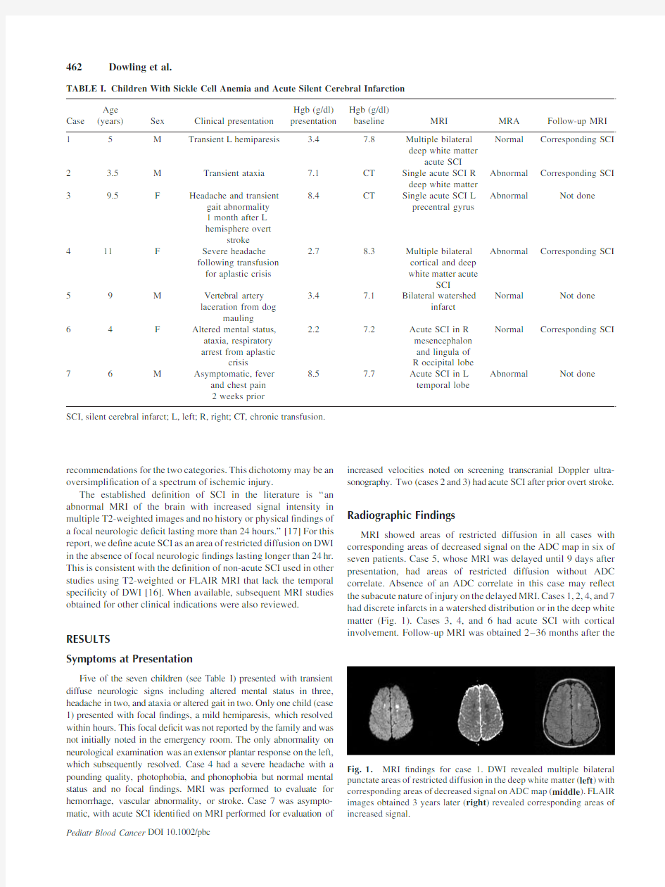
MRI showed areas of restricted diffusion in all cases with corresponding areas of decreased signal on the ADC map in six of seven patients.Case 5,whose MRI was delayed until 9days after presentation,had areas of restricted diffusion without ADC correlate.Absence of an ADC correlate in this case may re ?ect the subacute nature of injury on the delayed MRI.Cases 1,2,4,and 7had discrete infarcts in a watershed distribution or in the deep white matter (Fig.1).Cases 3,4,and 6had acute SCI with cortical involvement.Follow-up MRI was obtained 2–36months after the
Pediatr Blood Cancer DOI 10.1002/pbc
TABLE I.Children With Sickle Cell Anemia and Acute Silent Cerebral Infarction Case Age (years)Sex Clinical presentation Hgb (g/dl)presentation
Hgb (g/dl)baseline MRI MRA Follow-up MRI 15M Transient L hemiparesis
3.47.8Multiple bilateral deep white matter
acute SCI Normal Corresponding SCI 2 3.5M Transient ataxia 7.1CT Single acute SCI R deep white matter Abnormal Corresponding SCI
3
9.5
F
Headache and transient
gait abnormality 1month after L hemisphere overt
stroke 8.4
CT
Single acute SCI L precentral gyrus
Abnormal
Not done
411F Severe headache following transfusion for aplastic crisis 2.78.3
Multiple bilateral cortical and deep white matter acute
SCI
Abnormal Corresponding SCI
59M Vertebral artery laceration from dog
mauling
3.47.1Bilateral watershed
infarct Normal Not done 6
4
F
Altered mental status,ataxia,respiratory arrest from aplastic
crisis
2.2
7.2
Acute SCI in R mesencephalon and lingula of R occipital lobe Normal
Corresponding SCI
76M Asymptomatic,fever
and chest pain 2weeks prior
8.57.7Acute SCI in L temporal lobe
Abnormal Not done
SCI,silent cerebral infarct;L,left;R,right;CT,chronic
transfusion.
Fig.1.MRI ?ndings for case 1.DWI revealed multiple bilateral punctate areas of restricted diffusion in the deep white matter (left )with corresponding areas of decreased signal on ADC map (middle ).FLAIR images obtained 3years later (right )revealed corresponding areas of increased signal.
462Dowling et al.
acute SCI for cases1,2,4,and6.All four had FLAIR abnormalities corresponding to the areas of prior restricted diffusion indicating permanent neurologic injury.
Clinical Setting
Four of the acute SCI occurred in the clinical setting of an acute anemic event,that is,an exacerbation of the otherwise stable chronic anemia of HbSS.Case5presented with blood loss after a traumatic arterial laceration to a hemoglobin concentration of3.4g/dl.Cases 1,4,and6presented with acute anemic events more common in patients with HbSS,that is,aplastic crisis,with nadir hemoglobin concentrations between2.2and3.4g/dl.Their imaging studies revealed discrete or punctate unilateral or bilateral lesions,in a pattern typical of that observed in remote SCI in children with HbSS.Magnetic resonance angiography(MRA)was normal in three of these four cases associated with acute anemic events.Case3 had acute SCI affecting the cortex,1month after a larger left hemisphere overt stroke and may represent infarction of an adjacent area of brain with tenuous blood supply.
DISCUSSION
These observations suggest that SCI are detectible in the acute phase by DWI,that SCI onset may be associated with subtle, transient neurologic signs and symptoms,and that these acute SCI represent permanent brain injuries,not transient reversible changes or artifact.The presence of lesions typical for SCI on the follow-up imaging studies corresponding to the areas of restricted diffusion con?rms a fundamental assumption about SCI:they do,in fact, represent ischemic lesions.They are not due to some other pathologic process such as demyelination,which would have a different pattern of evolution of signal characteristics on MRI.
The temporal quality of the DWI signal changes may assist in establishing a relationship between concurrent clinical events and the acute SCI.Our observation of acute SCI in the clinical setting of acute anemic events in four of seven cases suggests that accelerated anemia may be an additional risk factor for SCI.In HbSS,the risk of overt stroke is increased in severe anemia,either acutely proximate to the stroke or in the steady state[12].The risk of overt stroke following an acute anemic event due to parvovirus B19infection was58times greater than expected,suggesting a causal association[18].A prior report of acute SCI in HbSS also occurred in the setting of an acute anemic event[19].That patient was a5-year-old male who presented with a hemoglobin concentration of2g/dl.His MRI was similar to our case1,with punctate areas of increased signal on DWI.We suspect that the rate of unrecognized acute SCI during acute anemic events could be similarly increased.Such events could lead to small areas of inadequate perfusion of brain tissue within the watershed distribution and result in small,clinically silent infarctions.
An increased index of suspicion for TIA or stroke is warranted for children with HbSS and increased education about stroke warning signs for patients and families could lead to increased and earlier diagnosis.Detection of these acute SCI has led to further evaluation and has altered the clinical management of several of these children.Increased vigilance for acute SCI in children with HbSS who present with subtle,transient neurologic symptoms is warranted.Unfortunately,the incidence of acute SCI cannot be determined from a retrospective review of our patients,because our clinical index of suspicion has increased over time with the identi?cation of these cases.Prospective studies are needed to detect acute SCI in the appropriate clinical settings,such as acute anemic events and acute chest syndrome,which are known risk factors for overt stroke in HbSS.A better understanding of the etiologies of SCI in the population at highest risk,children with HbSS,may also provide insight into the problem of SCI in the general population. This might allow for the prevention or the amelioration of the neuro-cognitive sequelae of these not-so-silent strokes. ACKNOWLEDGMENT
The authors were supported by the NHLBI Comprehensive Sickle Cell Center Program U54HL70588.M.M.D.and C.T.Q. were supported by the North and Central Texas Clinical and Translational Science Initiative from the NIH KL2RR024983,and M.M.D.is supported by the First American Real Estate Information Services,Inc.We thank John D.Nelson,MD and Michael R. DeBaun,MD,MPH for their valuable input. REFERENCES
1.Steen RG,Emudianughe T,Hankins GM,et al.Brain imaging
?ndings in pediatric patients with sickle cell disease.Radiology 2003;228:216–225.
2.DeBaun MR,Schatz J,Siegel MJ,et al.Cognitive screening
examinations for silent cerebral infarcts in sickle cell disease.
Neurology1998;50:1678–1682.
3.Schatz J,Brown RT,Pascual JM,et al.Poor school and cognitive
functioning with silent cerebral infarcts and sickle cell disease.
Neurology2001;56:1109–1111.
4.White DA,Moinuddin A,McKinstry RC,et al.Cognitive screening
for silent cerebral infarction in children with sickle cell disease.J Pediatr Hematol Oncol2006;28:166–169.
https://www.360docs.net/doc/c4463510.html,ler ST,Macklin EA,Pegelow CH,et al.Silent infarction as a
risk factor for overt stroke in children with sickle cell anemia:A report from the Cooperative Study of Sickle cell disease.J Pediatr 2001;139:385–390.
6.Mercuri E,Faundez JC,Roberts I,et al.Neurological‘soft’signs
may identify children with sickle cell disease who are at risk for stroke.Eur J Pediatr1995;154:150–156.
7.DeBaun MR,Derdeyn CP,McKinstry RC.Etiology of strokes in
children with sickle cell anemia.Ment Retard Dev Disabil Res Rev 2006;12:192–199.
8.Kinney TR,Sleeper LA,Wang WC,et al.Silent cerebral infarcts in
sickle cell anemia:A risk factor analysis.Pediatrics 1999;103:640–645.
9.Ohene-Frempong K,Weiner SJ,Sleeper LA,et al.Cerebrovascular
accidents in sickle cell disease:Rates and risk factors.Blood 1998;91:288–294.
10.Adams R,McKie V,Nichols F,et al.The use of transcranial
ultrasonography to predict stroke in sickle cell disease.N Engl J Med1992;326:605–610.
11.Balkaran B,Char G,Morris JS,et al.Stroke in a cohort of patients
with homozygous sickle cell disease.J Pediatr1992;120:360–366.
12.Quinn CT,Miller ST.Risk factors and prediction of outcomes in
children and adolescents who have sickle cell anemia.Hematol Oncol Clin N Am2004;18:1339–1354.
13.Muir KW,Buchan A,von Kummer R,et al.Imaging of acute stroke.
Lancet Neurol2006;5:755–768.
Pediatr Blood Cancer DOI10.1002/pbc
Acute Silent Infarction in HbSS463
14.Das RR,Seshadri S,Beiser AS,et al.Prevalence and correlates of
silent cerebral infarcts in the Framingham Offspring Study.Stroke 2008;39:2929–2935.
15.Oppenheim C,Lamy C,Touze E,et al.Do transient ischemic
attacks with diffusion-weighted imaging abnormalities correspond to brain infarctions?Am J Neuroradiol2006;27:1782–1787. 16.Kirkham FJ,Lerner NB,Noetzel M,et al.Trials in sickle cell
disease.Pediatr Neurol2006;34:450–458.17.Buchanan GR,DeBaun MR,Quinn CT,et al.Sickle cell
disease.Hematology(Am Soc Hematolo Educ Program)2004;
35–47.
18.Wierenga KJ,Serjeant GR.Cerebrovascular complications and
parvovirus infection in homozygous sickle cell disease.J Pediatr 2001;139:438–442.
19.Zimmerman RA.MRI/MRA evaluation of sickle cell disease of the
brain.Pediatr Radiol2005;35:249–257.
Pediatr Blood Cancer DOI10.1002/pbc 464Dowling et al.