Relation of body composition, fat mass, and serum lipids to
Relation of body composition,fat mass,and serum lipids to
osteoporotic fractures and bone mineral density in Chinese men and women 1–3
Yi-Hsiang Hsu,Scott A Venners,Henry A Terwedow,Yan Feng,Tianhua Niu,Zhiping Li,Nan Laird,Joseph D Brain,Steve R Cummings,Mary L Bouxsein,Cliff J Rosen,and Xiping Xu
ABSTRACT
Background:Higher fat mass may be an independent risk factor for osteoporosis and osteoporotic fractures.
Objective:We aimed to determine the independent contribution of fat mass to osteoporosis and to estimate the risk of osteoporotic fractures in relation to body weight,lean mass,and other confound-ers.
Design:This was a community-based,cross-sectional study of 7137men,4585premenopausal women,and 2248postmenopausal women aged 25–64y.Total-body and hip bone mineral content (BMC)and bone mineral density (BMD)and body composition were measured by dual-energy X-ray absorptiometry.Serum lipids were measured.Sex-and menopause-specific multiple generalized linear models were applied.
Results:Across 5-kg strata of body weight,fat mass was signifi-cantly inversely associated with BMC in the whole body and total hip.When we compared the highest quartile with the lowest quartile of percentage fat mass in men,premenopausal women,and post-menopausal women,the adjusted odds ratios (95%CIs)of osteopo-rosis defined by hip BMD were 5.2(2.1,13.2),5.0(1.7,15.1),and 6.9(4.3,11.2),respectively.Significant linear trends existed for higher risks of osteoporosis,osteopenia,and nonspine fractures with higher percentage fat mass.Significant negative relations were found be-tween whole-body BMC and cholesterol,triacylglycerol,LDL,and the ratio of HDL to LDL in all groups.
Conclusions:Risks of osteoporosis,osteopenia,and nonspine frac-tures were significantly higher for subjects with higher percentage body fat independent of body weight,physical activity,and age.Thus,fat mass has a negative effect on bone mass in contrast with the positive effect of weight-bearing itself.Am J Clin Nutr 2006;83:146–54.
KEY WORDS Bone mineral density,osteoporosis,fracture,body composition,lipids INTRODUCTION
Osteoporosis and its related fractures have become a major problem in elderly populations.Body weight is one of the stron-gest positive predictors of bone mass.A positive association between body weight and bone mass in subjects of all age groups has been shown (1–3).Higher body weight is thought to affect bone mass by increasing the mechanical stress mediated through muscle or by mass gravitational action through load placed on the
skeleton,thereby increasing the stimulus for osteogenesis (4,5).Lean,fat,and bone mass are the 3components of body weight.Lean and fat mass together account for 95%of body weight.Several epidemiologic studies have reported that both fat mass and lean mass may help to determine bone mass (6–13).An association between lean mass and bone mass may be due to mechanical load forces on bone.Because fat tissue is metaboli-cally active,its effect on the skeleton may be influenced not only by the weight-bearing effect but also by other non-weight-bearing effects,including the hormonal metabolism of adipo-cytes.Because of strong collinearity between fat mass and body weight,most epidemiologic studies that had small sample sizes could not explore the effects of fat mass on bone mass indepen-dent of body weight.
In contrast with epidemiologic studies,animal and in vitro studies support a negative effect of fat mass on bone mass.A possible link between fat tissue and bone tissue is the common stromal cell origin of both osteoblasts and adipocytes (14,15).Stromal cells in the marrow can differentiate into one of several mature forms,including osteoblasts and adipocytes.Under in vitro conditions,bone loss is associated with an expansion of adipose tissue in the marrow (16).A recent study showed that the peroxisome proliferator-activated receptor ?pathway,the dom-inant regulator of adipogenesis,not only determines adipocyte differentiation from mesenchymal progenitors,but also inhibits
1
From the Program for Population Genetics,Harvard School of Public Health,Boston,MA (Y-HH,SAV,HAT,YF,and JDB);the Division of Preventive Medicine,Department of Medicine,Brigham and Women Hos-pital,Harvard Medical School,Boston,MA (TN);Anhui Medical Univer-sity,Institute of Medicine,Anhui,China (ZL and XX);the Department of Biostatistics,Harvard School of Public Health,Boston,MA (NL);the San Francisco Coordinating Center,University of California,San Francisco,CA (SRC);the Beth Israel Deaconess Medical Center,Boston,MA (MLB);the Maine Center for Osteoporosis Research and Education,St Joseph Hospital,Bangor,ME (CJR);and the Center for Population Genetics,School of Public Health,University of Illinois at Chicago (XX).2
Supported by National Institute of Arthritis and Musculoskeletal and Skin Diseases grant R01AR045651.3
Address reprint requests to X Xu,Center for Population Genetics,School of Public Health,University of Illinois at Chicago,1603West Taylor Street,Room 978A,Chicago,IL 60612.E-mail:xipingxu@https://www.360docs.net/doc/f08530598.html,.E-mail:xipingxu@https://www.360docs.net/doc/f08530598.html,.
Received March 17,2005.
Accepted for publication August 25,2005.
146
Am J Clin Nutr 2006;83:146–54.Printed in USA.?2006American Society for Nutrition
by guest on December 13, 2012
https://www.360docs.net/doc/f08530598.html, Downloaded from C1.html
https://www.360docs.net/doc/f08530598.html,/content/suppl/2006/01/25/83.1.146.D
Supplemental Material can be found at:
osteoblast differentiation (17).Other evidence for a link between fat and bone mass is the action of leptin.Leptin is an anorexigenic metabolic hormone that is secreted in proportion to fat mass (18).Leptin’s anti-osteogenic action was demonstrated by intracere-broventricular injection of leptin into an ob/ob mouse,which decreased bone formation (19).A strong positive correlation between fat mass and serum lipid concentrations has been re-ported (20).Studies have shown that hyperlipidemia may con-tribute to osteoporosis by increasing osteoclastic bone resorption (21)and osteoclast viability (22).All of the above imply an inverse reciprocal relation between fat mass and bone mass.In the present study,we investigated the association between bone mass and body fat composition for a given body weight and estimated the risk of higher percentage fat mass (%FM)on os-teoporosis,osteopenia,and nonspine fractures by adjusting for body weight and other possible confounders in a large-scale cohort of men,premenopausal women,and postmenopausal women.
SUBJECTS AND METHODS
Study population
This study is part of an ongoing community-based osteoporo-sis study initiated in 2003among residents of Anhui Province,China.Men and women aged 25–64y were recruited.Partici-pants with a history of the following conditions were excluded from the study:type 1diabetes;renal failure;chronic infections,such as tuberculosis or other diseases;malignancy;rickets or other metabolic bone diseases;chronic glucocorticoid use;viral cirrhosis;and thyrotoxicosis.
This study was approved by the Human Subjects Committee (the institutional review board)of the Harvard School of Public Health and the Ethics Committee of Anhui Medical University.Written informed consent was obtained from each participant.Measurement of bone mineral content,bone mineral density,and body composition
Dual-energy X-ray absorptiometry (GE-lunar Prodigy,Waukesha,WI)was used to measure soft-tissue body composi-tion,bone mineral content (BMC,in g),and bone mineral density (BMD,in g/cm 2)through whole-body and total-hip scans.Whole-body fat mass and lean mass were expressed in terms of weight (g)and as a percentage of body weight.We define osteo-porosis as a total-hip BMD of 2.5SDs below the average peak BMD of young,healthy Chinese in same study area between 25and 30y of age (T-score ?2.5).Osteopenia was defined as a total-hip BMD between 1and 2.5SDs below the peak BMD (?2.5 T-score ?1).Anthropometry
A general physical examination was conducted of each par-ticipant.Height (m)was measured to the nearest 0.1cm on a portable stand meter,and weight was measured to the nearest 0.1kg with the subject standing motionless in the center of the scale.Weight and height were measured without the subjects’wearing shoes.Body mass index (BMI)was calculated as weight/height 2.Serum lipid measurements
Fasting blood samples were collected and stored in aliquots at ?80°C.Serum cholesterol was measured enzymatically with a
Cobas Integra Roche analyzer (Roche,Indianapolis,IN).Serum triacylglycerol was assayed by using the glyceryl dehydrogenase reaction after enzymatic hydrolysis of the glycerides on the Cobas Integra Roche analyzer.HDL cholesterol was measured after precipitation of LDL and VLDL with polyanions and phos-photungstic acid–magnesium chloride.The supernatant portion was assayed enzymatically on the Cobas Integra Roche analyzer.Questionnaires
Comprehensive questionnaires were used to collect the par-ticipants’demographic,occupational,and lifestyle information;reproductive history;disease history;consumption of alcohol;cigarette smoking;physical activity;history of fractures;and daily diet.A fracture questionnaire was applied for those partic-ipants who self-reported their fracture history.Fracture sites,treatments,and the age of the participants when they had the fractures were recorded.For this analysis,a nonspine fracture case was defined as participants with fractures at nonspine sites that occurred within 2y of the BMD measurements.
Statistical analyses
Participants were divided into men,premenopausal women,and postmenopausal women.We defined menopausal status by questionnaire.Because sex and menopausal status are 2of the most important predictors of bone mass,osteoporosis,and frac-tures,we report analyses separately on the basis of sex and menopausal status.The SAS 8.2software package (SAS Institute Inc,Cary,NC)was used to perform all statistical analyses.Univariate analyses
Analysis of variance (ANOVA)tests and chi-square tests were used to compare the principal characteristics of the study subjects among sex and menopausal status groups.Tukey’s test was also used to perform pairwise comparisons among groups when there was significance for an ANOVA or chi-square test.We then further divided %FM into quartiles.For covariates among quar-tiles of %FM,the generalized linear model was used to test for linear trend.Spearman’s rank correlation coefficients were used to determine the strength of relations between fat mass (or lean mass)and other variables,such as weight,serum lipid profile,and physical activity.t Tests were used to test the significance of the correlations.Multivariate analyses
To assess the effects of fat mass on BMC independently from the effects of body weight,quartiles of fat mass in 5-kg strata of weight were plotted against BMC.For maximum statistical power,only strata with ?200persons were included.Multiple linear regression models adjusted for differences in age,height,occupation,cigarette smoking (for men only),alcohol consump-tion (for men only),physical activity,and years since menopause (for postmenopausal women only)were used in each stratum to test for a linear trend in the relation of fat mass to BMC.Least-squares means and SDs of BMC were computed.Multivariate logistic regression models adjusted for age,physical activity,occupation,cigarette smoking,alcohol consumption,height,weight,and years since menopause (for postmenopausal women only)were used to estimate the independent risks of %FM on osteoporosis and osteopenia.For the risk of %FM on nonspine
BODY COMPOSITION AND BMD IN A CHINESE POPULATION 147
by guest on December 13, 2012
https://www.360docs.net/doc/f08530598.html,
Downloaded from
fractures,the adjusted variables were age,physical activity,oc-cupation,whole-body BMD,and years since menopause (for postmenopausal women only).The log likelihood ratio test was used to test the interaction among covariates.Linear regression models adjusted for age,height,%FM,occupation,physical ac-tivity,cigarette smoking,alcohol consumption,and years since menopause (for postmenopausal women only)were applied to explore the magnitude of relations between serum lipid profiles (cholesterol,triacylglycerol,HDL,LDL,and the ratio of LDL to HDL)and bone mass (BMD and BMC).Generalized estimating equation models were used to adjust for intraclass correlation within family members.
RESULTS
Principal and clinical characteristics
The analyses included 13970subjects (7137men,4585pre-menopausal women,and 2248postmenopausal women).The principal characteristics of the study population,including the distributions of the serum lipids profile,body composition,BMD,and BMC,are shown in Table 1.Significant differences in all reported covariates were found among the 3groups.Age,weight,height,BMI,BMD,BMC,body composition,cigarette smoking,and secondhand smoking status differed significantly among the 3groups (P 0.01).Men had significantly higher percentages of alcohol consumption and heavy physical activity and a lower percentage of occupation as a farmer than both
premenopausal and postmenopausal women.No significant dif-ferences were found in alcohol consumption,physical activity,or occupation between premenopausal and postmenopausal women.As shown in the table,our study population was rela-tively lean,especially compared with populations in North America or Europe.
Negative associations between bone mineral content and fat mass
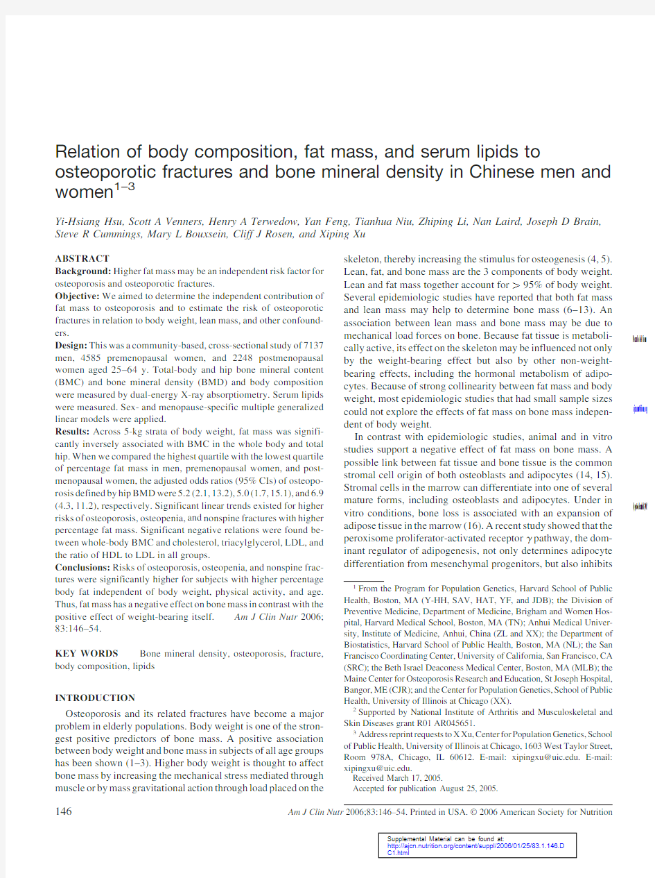
We further divided the subjects into 5-kg strata of body weight.As shown in Figure 1,there were 7weight strata (from 45to 80kg)in men and 6weight strata (from 40to 69kg)in premeno-pausal and postmenopausal women.The least-squares means and SDs of total-hip BMC in each quartile of fat mass among weight strata were plotted.A significant fat mass ?weight ?group interaction for total-hip BMC (P 0.0001)was found by log likelihood ratio test.Significant fat mass ?weight interac-tions were also found in men (P 0.0001),premenopausal women (P ?0.01),and postmenopausal women (P ?0.005).Fat mass explained 0.5%to 24%of BMC variation throughout body weight strata.Significant negative associations between fat mass and BMC at the total hip were found in all the weight strata.A significant fat mass ?weight ?group interaction for whole-body BMC (P 0.0001)was found by log likelihood ratio tests.Significant fat mass ?weight interactions were also found in men (P 0.0001),premenopausal women (P ?0.001),and postmenopausal women (P ?0.01).We also found significant
TABLE 1
Principal characteristics of the study population stratified by sex and menopausal status 1
Variable
Men (n ?7137)Premenopausal
women (n ?4585)Postmenopausal
women (n ?2248)Age (y)47.3?7.7b,241.5?5.3a 52.6?4.7c Weight (kg)57.5?8.1c 53.1?7.5b 51.1?7.7a Height (cm)163.5?5.8c 153.9?5.0b 152.6?5.1a BMI (kg/m 2)
21.4?2.5a
22.4?2.8c
21.9?2.9b Age at menopause (y)46.4?4.8Time since menopause (y)NA NA 6.2?5.5Bone mineral density (g/cm 2)Whole body 1.13?0.08c 1.11?0.07b 1.02?0.09a Total hip
0.98?0.12c 0.97?0.11b 0.86?0.12a Bone mineral content (g)Whole body 2546.2?345.1c
2190.3?290.7b
1906.7?315.7a
Total hip
34.1?5.4c
28.9?4.1b
25.7?6.4a
Body composition Fat mass (kg)7.9?5.1a 15.4?5.3c 14.6?5.5b Lean mass (kg)
46.7?6.0c 37.7?4.3b 36.6?4.5a Percentage fat mass (%)12.1?6.2a 25.9?6.4c 25.2?6.7b Percentage lean mass (%)83.3?6.3c 69.5?6.1a 70.5?6.5b Lifestyle [n (%)]Cigarette smoking 4986(70.1)c 85(1.9)a 117(5.2)b Secondhand smoking 3973(67.3)c 2592(60.0)b 976(46.3)a Alcohol consumption 3328(46.8)b 114(2.5)a 84(3.7)a Heavy physical activity 42487(35.0)b 1508(33.0)a 703(31.4)a Occupation as a farmer
5853(82.4)a
4092(89.6)b
2015(90.1)b
1
NA,not applicable.Means in a row with different superscript letters are significantly different,P 0.05(ANOVA and Tukey’s test for continuous variables and chi-square test for categorical variables).
2x ??SD (all such values).3
Secondhand smoking among non-cigarette-smokers.4
More than 5h/d carrying (on back or shoulder),raising,lifting,or moving a load of ?20kg.
148HSU ET AL
by guest on December 13, 2012
https://www.360docs.net/doc/f08530598.html,
Downloaded from
negative associations between fat mass and whole-body BMC (see Figure 1under “Supplemental data”in the current issue at https://www.360docs.net/doc/f08530598.html,).
Percentage fat mass and odds ratios of osteoporosis,osteopenia,and nonspine fracture
We then divided the subjects into quartiles of %FM.The average %FM in each quartile of %FM is shown in Table 2.
Participants with higher %FM tended to have lower percentage lean mass (%LM);lower physical activity;higher body weight;higher cholesterol,triacylglycerol,and LDL concentrations;lower HDL concentrations;and a higher ratio of LDL to HDL.Significant interactions were found between quartiles of %FM and sex for %LM,weight,serum lipids,and physical activity by log likelihood ratio test (P values 0.001).
We defined 156(2.2%)men,82(1.8%)premenopausal women,and 278(12.4%)postmenopausal women as having osteoporosis.The numbers of osteoporosis,osteopenia,and non-spine fracture subjects in each quartile of %FM are shown in Table 3.Significant linear trends of higher ORs of osteoporosis,osteopenia,and nonspine fracture with higher %FM were found (P 0.0001,P 0.0001,and P ?0.002,respectively)in mul-tiple logistic regression models that included all subjects and that were adjusted for body weight and other covariates.A significant group ?quartile of %FM interaction for the OR of osteoporosis was found (P ?0.02),but not for the ORs of osteopenia or fracture (P ?0.3and 0.2,respectively).As shown in Table 3,comparing the highest quartile (Q4)with the lowest quartile of %FM,the adjusted ORs (95%CI)of osteoporosis were 5.2(2.1,13.2),5.0(1.7,15.1),and 6.9(4.3,11.2),in men,premenopausal women,and postmenopausal women,respectively.
A total of 1823(25.6%)men,1172(25.6%)premenopausal women,and 1166(51.9%)postmenopausal women were defined as having osteopenia.There were significant and independent linear trends for higher ORs of osteopenia with higher %FM (P values 0.01in all groups).
A total of 82(1.1%)men,54(1.2%)premenopausal women,and 46(2.0%)postmenopausal women self-reported at least one nonspine fracture.There were significant and independent linear trends for higher ORs of nonspine fractures in men and premeno-pausal women with higher %FM (P values 0.05in men and premenopausal women).
Relative odds by lean mass,physical activity,and years since menopause
We investigated the ORs of osteoporosis,osteopenia,and non-spine fracture by %LM,physical activity,and years since meno-pause.Weight was positively correlated with both fat mass and lean mass (see Table 1under “Supplemental data”in the current issue at https://www.360docs.net/doc/f08530598.html,).Participants with higher weights tended to have a higher %FM and a lower %LM,and participants with lower weights had lower %FM and higher %LM.The crude and adjusted risks of %LM,heavy physical activity,and years since menopause for osteoporosis,osteopenia,and nonspine fractures for all subjects are shown in Table 4.These covariates have been reported as important risk factors that may influence BMD and cause osteoporosis.Heavy physical activity reduced the risk of osteoporosis and osteopenia but increased the risk of nonspine fractures after adjustment for other covariates.Years since meno-pause increased the odds of osteoporosis and osteopenia.We divided %LM into quartiles.The mean (?SD)values for Q1to Q4were 64.8?3.53%,72.9?1.95%,80.8?2.61%,and 88.6?1.74%,respectively.%LM slightly decreased the ad-justed ORs of osteopenia and fractures,but significantly so only for osteopenia.We used the generalized linear model to test for linear trends.After adjustment for possible confounders,there were significant linear trends for osteopenia (P 0.01)
and
FIGURE 1.Least-squares mean (?SD)total-hip bone mineral content (BMC)stratified by fat mass in 5-kg strata of body weight in men,premeno-pausal women,and postmenopausal women.The bars from left to right are quartiles (Q)1,2,3,and 4of percentage fat mass in each body weight stratum.Multiple linear regression models adjusted for age,height,occupation,cig-arette smoking (for men only),alcohol consumption (for men only),physical activity,and years since menopause (for postmenopausal women only)were used to test for linear trends in total-hip BMC by percentage fat mass quartiles within weight strata.*P 0.05;**P 0.01;***P 0.001(Wald chi-square test).P 0.0001for fat mass ?weight ?group interaction;fat mass ?weight interactions were also found in men (P 0.0001),premenopausal women (P ?0.01),and postmenopausal women (P ?0.005).
BODY COMPOSITION AND BMD IN A CHINESE POPULATION 149
by guest on December 13, 2012
https://www.360docs.net/doc/f08530598.html,
Downloaded from
nonspine fracture (P 0.05),but not for osteoporosis (P ?0.8),with increasing %LM.There were neither significant interac-tions between groups and %LM (P ?0.2,0.1,and 0.4,respec-tively,for osteoporosis,osteopenia,and fracture)nor between groups and physical activity (P ?0.8,0.1,and 0.7,respectively,for osteoporosis,osteopenia,and fracture).Interaction of %FM and heavy physical activity
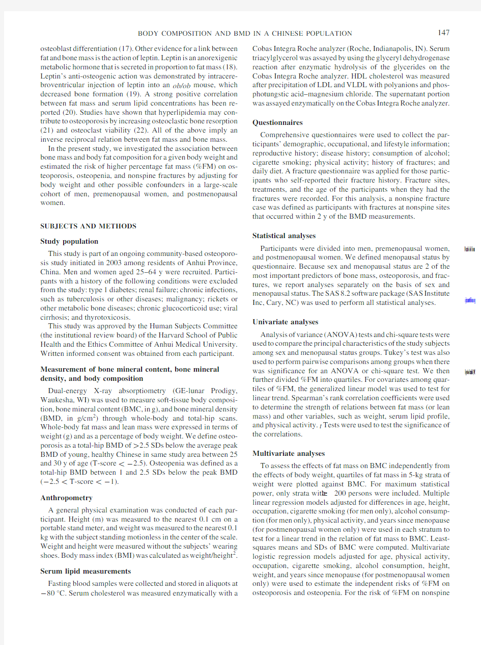
Because physical activity is negatively correlated with %FM (see Table 1under “Supplemental data”in the current issue at https://www.360docs.net/doc/f08530598.html,),we estimated the interaction of %FM and heavy physical activity for the ORs of osteoporosis and osteopenia.A significant 3-way interaction (group ?%FM ?physical activity)for risk of osteoporosis was found (P 0.001),but not for risk of osteopenia (P ?0.4).There was neither a significant interaction between %FM and physical activity for the odds of osteoporosis (P ?0.8,0.08,and 0.3,respectively,for men,premenopausal women,and postmeno-pausal women)nor a significant interaction between %FM and physical activity for the odds of osteopenia (P ?0.3,0.1,and 0.4,respectively,for men,premenopausal women,and postmenopausal women).As shown in Figure 2,a linear trend toward higher ORs of osteoporosisandosteopeniawithhigher%FMwasobservedwithor without heavy physical activity.For osteoporosis,the adjusted ORs of Q4and Q3compared with the lowest quartile in premenopausal
TABLE 2
Mean percentage fat mass (%FM)and other covariates by quartiles (Q)of %FM 1Variable
Q1Q2Q3Q4Men n 1784178417841784%FM 5.7?0.828.7?1.013.0?1.521.1?4.0%LM 3,4
90.0?1.086.9?1.182.2?1.874.3?3.7Weight (kg)3,451.7?5.254.6?5.257.9?5.865.6?8.1Age (y)5
47.8?7.747.6?7.647.3?7.746.5?7.6Cholesterol (mg/dL)3,4
161.9?29.4165.7?30.5171.3?30.6180.7?33.1Triacylglycerol (mg/dL)3,435.1?14.637.9?16.043.7?21.860.9?31.7HDL (mg/dL)3,453.0?12.852.4?13.051.4?13.146.1?11.2LDL (mg/dL)3,492.4?22.095.3?23.098.8?23.8104.3?27.2LDL:HDL 3,4
1.8?0.5 1.9?1.6
2.0?0.6 2.3?0.7Occupation as a farmer [n (%)]6
1643(92)1541(87)1434(81)1234(69)Heavy physical activity [n (%)]3,4,7769(43)696(39)616(35)406(23)Premenopausal women n 1146114611461146%FM 18.0?2.723.5?1.227.9?1.534.4?2.9%LM 3,4
77.1?3.071.4?1.767.5?1.661.8?2.8Weight (kg)3,447.2?5.151.8?5.854.4?6.658.9?6.9Age (y)5
41.6?5.341.6?5.341.3?5.241.5?5.4Cholesterol (mg/dL)3,4
159.2?28.5163.9?30.4166.0?30.8171.5?31.7Triacylglycerol (mg/dL)3,442.0?17.747.2?20.949.3?22.956.7?26.8HDL (mg/dL)3,450.1?10.749.1?11.348.2?10.946.6?10.7LDL (mg/dL)3,489.7?22.592.9?24.894.8?24.298.1?25.4LDL:HDL 3,4
1.8?0.5 1.9?0.6
2.0?0.6 2.2?0.6Occupation as a farmer [n (%)]6
1058(93)1003(88)1006(88)1024(90)Heavy physical activity [n (%)]3,4,7427(38)398(35)362(32)320(28)Postmenopausal women n 562562562562%FM 16.8?2.822.8?1.327.3?1.434.1?3.1%LM 3,4
78.9?3.272.5?1.768.3?1.862.4?3.0Weight (kg)3,444.5?4.649.7?5.353.0?7.357.2?6.9Age (y)5
52.6?4.952.5?4.752.5?4.752.5?4.7Cholesterol (mg/dL)3,4
174.9?31.9178.3?31.9182.3?32.6190.2?34.0Triacylglycerol (mg/dL)3,447.3?19.353.2?24.956.6?24.364.5?31.7HDL (mg/dL)3,853.8?12.152.1?11.951.5?11.950.4?11.4LDL (mg/dL)3,499.1?25.3101.5?26.1104.2?27.0108.6?26.9LDL:HDL 3,4
1.9?0.5
2.0?0.6 2.1?0.6 2.2?0.6Occupation as a farmer [n (%)]6
508(91)492(88)491(88)523(94)Heavy physical activity [n (%)]3,4,7
212(38)
167(30)
180(32)
143(26)
1%LM,percentage lean mass.2
x ??SD (all such values).3
Significant %FM ?group (sex and menopausal status)interactions,P 0.05(likelihood ratio test).4,8
For covariates among quartiles of %FM,the generalized linear model was used to test for linear trend in men,premenopausal women,and postmeno-pausal women separately (Wald chi-square test):4P 0.001,8P 0.01.
5
Significant linear trend (P ?0.03)for age with higher quartiles of %FM in the analysis of all samples.6
Significant linear trend (P 0.0001)for occupation with higher quartiles of %FM in the analysis of all samples.7
More than 5h/d carrying (on back or shoulder),raising,lifting,or moving a load of ?20kg.
150HSU ET AL
by guest on December 13, 2012
https://www.360docs.net/doc/f08530598.html,
Downloaded from
women who performed heavy physical activity are not shown be-cause there were too few subjects ( 5).
Associations between bone mass and the serum lipid profile In addition to body weight and physical activity,serum lipids were significantly correlated with fat mass and lean mass.Cho-lesterol,triacylglycerol,and LDL concentrations and the ratio of LDL to HDL were positively correlated with fat mass,but HDL was negatively correlated with fat mass.The correlations be-tween serum lipids and fat mass were obviously higher than the correlations between serum lipids and lean mass (see Table 1under “Supplemental data”in the current issue at https://www.360docs.net/doc/f08530598.html, ).Adjusted linear regression models were applied to explore the magnitude of relations between serum lipid profiles and whole-body BMC (Table 5).Significantly negative associations were found between whole-body BMC and cholesterol,triacylglyc-erol,and LDL concentrations and the ratio of HDL to LDL.No significant associations between whole-body BMC and HDL were found.The same patterns of relations were found between total-hip BMC and serum lipids,but the associations were not significant.
DISCUSSION
Our study showed significantly higher ORs of osteoporosis,osteopenia defined by BMD,and nonspine fractures in subjects with higher %FM after adjustment for possible confounders.The higher ORs were associated with %FM independently of body weight,age,and physical activity.Furthermore,the large sample size allowed us to place the subjects into 5-kg strata of body weight and to still have enough statistical power to assess the
effect of fat mass on bone mass independently of body weight.Fat mass was negatively associated with BMC in the whole body and total hip for a given body weight.The negative associations between fat mass and BMC were independent of age and physical activity.Thus,fat mass seems to affect BMD and BMC beyond the effect of weight bearing itself.
Lean mass and fat mass together account for 95%of body weight.The remaining 5%is bone mass.Lean mass has been reported as a predictor of bone mass through its mechanical pull on the skeleton (2).%FM is an estimate of the proportion of body weight that is fat tissue.This means that %FM can differentiate characteristics of the tissue types from the mass effect of the tissues.In our study,%FM and %LM had a reciprocal relation.It is reasonable to doubt that the riskofhigher%FMonosteoporosismaybeduetotheeffectoflower %LM.As shown in Table 4,the ORs of osteoporosis were lower in subjects with higher %LM after adjustment for body weight and other confounders.Because the correlations between body weight and %LM were negative,the effect of %LM on osteoporosis and osteopenia without adjustment for body weight seemed only to reflect the effect of body weight.However,it was difficult to dif-ferentiate the effect of %FM from that of %LM in our cross-sectional design.
%FM could be a surrogate marker for lifestyle factors that are themselves negatively associated with BMD.Intervention stud-ies have shown that exercise is associated with higher bone den-sity and lower fat mass (23,24).However,when we examined the ORs of %FM on bone outcomes,whether or not we took physical activity into account did not appreciably change the results.In addition,we still observed positive associations between %FM and osteoporosis in subjects with or without heavy physical
TABLE 3
Adjusted odds ratios (ORs)and 95%CIs of osteoporosis,osteopenia,and nonspine fractures by quartile (Q)of percentage fat mass (%FM)in men,premenopausal women,and postmenopausal women 1
Quartile
%FM Osteoporosis
Osteopenia
Nonspine fracture Cases Adjusted OR 2,3Cases Adjusted OR 2,4Cases Adjusted OR 4,5Men %n n n Q1 5.7?0.864 1.0574 1.020 1.0Q28.7?1.043 1.7(0.9,3.1)6517 1.3(1.0,1.5)17 1.0(0.5,2.0)Q313.0?1.527 3.3(1.4,7.9)7407 1.6(1.2,2.1)23 1.7(0.9,3.2)Q4
21.1?4.022 5.2(2.1,13.2)8325 3.6(2.2,5.9)22 2.3(1.1,4.9)Premenopausal women Q118.0?2.745 1.0366 1.08 1.0Q223.5?1.222 1.4(0.8,2.6)313 1.5(1.2,2.0)10 1.3(0.5,3.5)Q327.9?1.510 3.5(1.5,7.7)7250 1.7(1.2,2.5)15 2.1(0.8,5.1)Q4
34.4?2.95 5.0(1.7,15.1)7243 3.3(2.0,5.4)21 3.0(1.1,7.8)Postmenopausal women Q116.8?2.8117 1.0299 1.010 1.0Q222.8?1.363 2.6(1.7,3.9)7303 1.4(0.9,2.0)10 1.2(0.5,2.9)Q327.3?1.457 3.1(2.0,4.8)8299 2.1(1.3,3.5)15 1.7(0.7,4.3)Q4
34.1?3.1
41
6.9(4.3,11.2)8
265
2.6(1.3,5.5)
11
1.3(0.4,3.4)
1n ?1784,1146,and 562for each quartile in men,premenopausal women,and postmenopausal women,respectively.
2
Models were adjusted for age,physical activity,occupation,cigarette smoking,alcohol consumption,weight,height,and years since menopause (for postmenopausal women only).
3
Significant group ?quartile of %FM interaction for osteoporosis,P ?0.02.4
Significant linear trends of higher ORs of osteopenia and nonspine fracture with increased %FM in analysis including all subjects (P 0.0001and P ?0.002,respectively).
5
Models were adjusted for age,physical activity,occupation,whole-body bone mineral density,and weight.6–8
Wald chi-square test with Q1as the reference group in sex and menopause-specific multiple logistic regression models:60.05 P 0.09,7P 0.01,
8
P 0.001.
BODY COMPOSITION AND BMD IN A CHINESE POPULATION
151
by guest on December 13, 2012
https://www.360docs.net/doc/f08530598.html,
Downloaded from
activity.Therefore,the risks of %FM on osteoporosis,osteope-nia,and nonspine fractures were independent of physical activ-ity.Sex,menopausal status,and other environmental factors may confound the associations between fat mass and bone mass.Our large sample size allowed us to analyze men,premenopausal women,and postmenopausal women separately.Because of the limited availability of public transportation,the similarity of lifestyle,and the lack of calcium supplements or hormone re-placement therapy,the study population appeared to be relatively stable and fairly homogeneous.The potential for confounding in our study was at least significantly minimized.
Similar to previous studies,without control for body weight,we observed positive associations between fat mass and bone mass (6–13;see Table 2under “Supplemental data”in the cur-rent issue at https://www.360docs.net/doc/f08530598.html,).However,we found significantly negative relations between fat mass and bone mass for a given body weight.Other studies have shown the same phenomenon of fat mass being negatively correlated with bone mass (8,9,11)and forearm fractures (25)after control for body weight.As the result of strong collinearity between fat mass and weight,most other studies with small sample sizes could not reliably explore the effect of fat mass on BMD independently of body weight.
Fat mass likely affects bone mass through both weight-bearing and non-weight-bearing effects.Like lean mass,the weight-bearing effect is a positive one and is likely related to cortical and periosteal modeling.On the other hand,the non-weight-bearing effect of higher fat mass may be negative,particularly for this cohort.An animal study of the peroxisome proliferator-activated receptor ?pathway showed that the regulation of adipocyte dif-ferentiation may be important for the regulation of bone ho-meostasis (17).That study observed that peroxisome proliferator-activated receptor ?haploinsufficiency was shown to enhance osteoblastogenesis in vitro and to increase bone mass in mice at both 8and 52wk of age in vivo.This effect was not mediated by insulin or leptin.A positive correlation between fat mass and serum leptin concentrations was found in humans (8,11).Obese mouse models have shown that both leptin-deficient ob/ob mice and leptin-receptor-deficient db/db mice have
higher
FIGURE 2.Adjusted odds ratios (ORs)and 95%CIs of osteoporosis and osteopenia by percentage fat mass quartiles (Q)stratified by physical activity in men,premenopausal women,and postmenopausal women.Q1in each stratum is the reference group.Multiple logistic regression models adjusted for age,percentage fat mass,occupation,cigarette smoking,alcohol con-sumption,weight,height,and years since menopause (in postmenopausal women only)were used to estimate the ORs and 95%CIs.The Wald chi-square test was used to perform the significance test.The adjusted ORs of Q4and Q3for premenopausal women in the heavy physical activity stratum are not shown because there were too few subjects ( 5).A significant 3-way interaction (group ?percentage fat mass ?physical activity)for risk of osteoporosis was found (P 0.001).
TABLE 4
Odds ratios (ORs)and 95%CIs of osteoporosis,osteopenia,and nonspine fracture for percentage lean mass (%LM),physical activity,and years since menopause (postmenopausal women only)
Variables
Osteoporosis
Osteopenia
Nonspine fracture OR 1
Adjusted OR 2OR 1
Adjusted OR 1OR 1
Adjusted OR 1,3%LM 4
Q2 1.46(1.12,1.90)50.84(0.61,1.17) 1.26(1.13,1.39)60.90(0.80,1.03)0.93(0.63,1.37)0.87(0.56,1.34)Q3 1.63(1.26,2.10)60.91(0.61,1.37) 1.03(0.93,1.14)0.73(0.61,0.87)60.75(0.50,1.12)0.60(0.33,1.11)Q4
1.34(1.02,1.75)70.93(0.53,1.64) 1.23(1.11,1.36)50.74(0.60,0.92)50.67(0.44,1.02)0.46(0.21,1.00)8Heavy physical activity 0.73(0.61,0.90)50.74(0.59,0.93)70.93(0.86,1.01)80.87(0.80,0.94)5 1.23(0.91,1.66) 1.42(1.04,1.94)7Years since menopause
2.07(1.81,2.40)6
1.55(1.27,1.88)6
1.45(1.33,1.59)6
1.26(1.13,1.40)6
1.13(0.86,1.47)
1.08(0.77,1.52)
1Significant linear trend with increasing %LM,P 0.05.
2
%LM adjusted for sex,menopausal status,age,occupation,cigarette smoking,alcohol consumption,height,weight,and physical activity.Heavy physical activity adjusted for sex,menopausal status,age,occupation,cigarette smoking,alcohol consumption,height,weight,percentage fat mass,and years since menopause (postmenopausal women only).
3
%LM adjusted for sex,menopausal status,age,occupation,whole-body bone mineral density,weight,and physical activity.Heavy physical activity adjusted for sex,menopausal status,age,occupation,whole-body bone mineral density,weight,percentage fat mass,and years since menopause (postmeno-pausal women only).
4
The mean (?SDs)were 64.8?3.53%,72.9?1.95%,80.8?2.61%,and 88.6?1.74%for Q1to Q4,respectively.Q1of %LM was the reference group.Heavy physical activity adjusted for sex,menopausal status,age,occupation,cigarette smoking,alcohol consumption,height,weight,percentage fat mass,and years since menopause (postmenopausal women only).
5–8
Wald chi-square test in logistic regression models:5P 0.01,6P 0.001,7P 0.05,80.05 P 0.09.
152HSU ET AL
by guest on December 13, 2012
https://www.360docs.net/doc/f08530598.html,
Downloaded from
rates of bone formation,despite their hypogonadism and hyper-cortisolism,which reduce bone mass (26).However,the results of observational studies in humans are somewhat controversial.Both negative (11,13)and positive (8,9,27)associations be-tween serum leptin concentrations and bone mass have been reported.The observed positive correlations between leptin con-centrations and bone mass may be confounded by body weight (9,27)or by menopausal status (8).The above evidence seems to explain the negative effect of fat on bone formation.However,in addition to these hypotheses,other unknown factors may exist that link both fat tissue and bone tissue.These unknown factors require further investigation.
Besides weight and lean mass,serum lipids are strongly cor-related with fat mass.Epidemiologic evidence has linked osteo-porosis and cardiovascular disease (28).In our study,after ad-justment for weight,fat mass,and other confounders,significantly negative relations were found between whole-body BMC and cholesterol,triacylglycerol,and LDL concentrations.Similar patterns were also found between total-hip BMC and serum lipids,but these were not significant.Previous studies reported negative correlations between cholesterol,triacylglyc-erol,or LDL and bone mass at the spine and total body but not at the hip (29,30).A prospective study showed increasing serum cholesterol concentrations with decreasing BMD at the spine but not at the hip,independent of the change in BMI during an 8-y follow-up (29).After adjustment for BMI and age,a study with 1303postmenopausal women also reported a higher risk of os-teopenia for participants with higher plasma LDL concentrations (31).Studies have shown that oxidized lipids inhibit osteoblastic differentiation from preosteoblasts in vitro and bone formation in vivo.Products of lipoprotein oxidation inhibit preosteoblast dif-ferentiation (16,32)and result in reduced bone mineralization (33).Several studies indicated that statins,which are widely used as lipid-lowering agents,seem to provide benefits in the preven-tion of bone loss and fractures (34,35).Another study showed that lipid-lowering therapy results in slight increases in osteo-calcin without changes in collagen type I crosslinked carboxyl terminal peptide,which also suggests the interruption of serum lipids on osteoblast function (36).However,in our study,serum lipids may explain only a small part of the relation between fat mass and bone mass.
Compared with other studies,this population may represent a lean and underweight population.Because of the age inclusion criterion (25–64y old),the prevalence of osteoporosis in our study may not represent the prevalence in the general Asian population.The results from our study may also not apply di-rectly to populations other than Asians.Furthermore,our study,being observational in design,cannot prove the existence of a causal relation between fat mass and bone mass.Our cross-sectional design also cannot clarify whether fat mass or %FM was associated with peak bone density or with age-related bone loss.More studies are needed in other populations,particularly in those with higher BMIs,to explore this relation further.
In conclusion,the results of the present study show that body fat mass and serum lipids are negatively associated with bone mass for a given body weight in a relatively lean population.These results highlight the importance of %FM and serum lipid profiles as risk factors for osteoporosis and also provide a rationale for further exploration of the underlying
mechanisms.
We thank Melissa Veno for editing this manuscript.
Y-HH completed the data analyses and prepared the manuscript.SAV and MLB contributed to data analysis and manuscript preparation.HAT,CJR,SRC,and JDB participated in the study design and critically reviewed the manuscript.NL contributed to data analysis and critically reviewed the manuscript.YF,TN,ZL,and XX participated in the data collection.None of the authors had any financial or personal interest,including advisory board affiliations,in any company or organization sponsoring the research.
REFERENCES
1.Hannan MT,Felson DT,Anderson JJ.Bone mineral density in elderly men and women:results from the Framingham osteoporosis study.J Bone Miner Res 1992;7:547–53.
2.Edelstein SL,Barrett-Connor E.Relation between body size and bone mineral density in elderly men and women.Am J Epidemiol 1993;138:160–9.
3.Nguyen TV,Sambrook PN,Eisman JA.Bone loss,physical activity,and weight change in elderly women:the Dubbo Osteoporosis Epidemiology Study.J Bone Miner Res 1998;13:1458–67.
4.Beck TJ,Oreskovic TL,Stone KL,et al.Structural adaptation to chang-ing skeletal load in the progression toward hip fragility:the study of osteoporotic fractures.J Bone Miner Res 2001;16:1108–19.
https://www.360docs.net/doc/f08530598.html,nyon L,Skerry T.Postmenopausal osteoporosis as a failure of bone’s adaptation to functional loading:a hypothesis.J Bone Miner Res 2001;16:1937–47.
6.Reid IR,Ames R,Evans MC,et al.Determinants of total body and regional bone mineral density in normal postmenopausal women–a key role for fat mass.J Clin Endocrinol Metab 1992;75:45–51.
7.Reid IR,Plank LD,Evans MC.Fat mass is an important determinant of whole body bone density in premenopausal women but not in men.J Clin Endocrinol Metab 1992;75:779–82.
8.Pasco JA,Henry MJ,Kotowicz MA,et al.Serum leptin levels are associated with bone mass in nonobese women.J Clin Endocrinol Metab 2001;86:1884–7.
9.Yamauchi M,Sugimoto T,Yamaguchi T,et al.Plasma leptin concen-trations are associated with bone mineral density and the presence of vertebral fractures in postmenopausal women.Clin Endocrinol (Oxf)2001;55:341–7.
10.Wu F,Ames R,Clearwater J,Evans MC,Gamble G,Reid IR.Prospec-tive 10-year study of the determinants of bone density and bone loss in normal postmenopausal women,including the effect of hormone re-placement therapy.Clin Endocrinol (Oxf)2002;56:703–11.
11.Blum M,Harris SS,Must A,et al.Leptin,body composition and bone
mineral density in premenopausal women.Calcif Tissue Int 2003;73:27–32.
12.Capozza RF,Cointry GR,Cure-Ramirez P,Ferretti JL,Cure-Cure C.A
DXA study of muscle-bone relationships in the whole body and limbs of 2512normal men and pre-and post-menopausal women.Bone 2004;35:283–95.
TABLE 5
Regression coefficients (?)for serum lipids (mg/dL)to whole-body bone mineral content (BMC)1
Variable Whole-body BMC (g)Men Premenopausal
women Postmenopausal
women Cholesterol ?0.70?0.092?0.53?0.102?0.43?0.143Triacylglycerol ?0.99?0.142?1.12?0.142?0.55?0.183HDL 0.00?0.25?0.03?0.280.29?0.39LDL
?0.56?0.122?0.40?0.133?0.36?0.184LDL:HDL
?10.19?3.243
?13.95?5.574
?20.98?8.194
1
All values are ??SE.The regression models were adjusted for age,height,percentage fat mass,occupation,physical activity,cigarette smoking,alcohol consumption,and years since menopause (for postmenopausal women only).
2–4
Wald chi-square test in multiple linear regression models:2P 0.001,3
P 0.01,4P 0.05.
BODY COMPOSITION AND BMD IN A CHINESE POPULATION
153
by guest on December 13, 2012
https://www.360docs.net/doc/f08530598.html,
Downloaded from
13.Kontogianni MD,Dafni UG,Routsias JG,Skopouli FN.Blood leptin
and adiponectin as possible mediators of the relation between fat mass and BMD in perimenopausal women.J Bone Miner Res 2004;19:546–51.
14.Bruder SP,Fink DJ,Caplan AI.Mesenchymal stem cells in bone devel-opment,bone repair,and skeletal regeneration therapy.J Cell Biochem 1994;56:283–94.
15.Parhami F,Jackson SM,Tintut Y,et al.Atherogenic diet and minimally
oxidized low density lipoprotein inhibit osteogenic and promote adipo-genic differentiation of marrow stromal cells.J Bone Miner Res 1999;14:2067–78.
16.Parhami F,Morrow AD,Balucan J,et al.Lipid oxidation products have
opposite effects on calcifying vascular cell and bone cell differentiation.A possible explanation for the paradox of arterial calcification in osteo-porotic patients.Arterioscler Thromb Vasc Biol 1997;17:680–7.
17.Akune T,Ohba S,Kamekura S,et al.PPAR ?insufficiency enhances
osteogenesis through osteoblast formation from bone marrow progeni-tors.J Clin Invest 2004;113:846–55.
18.Auwerx J,Staels https://www.360docs.net/doc/f08530598.html,ncet 1998;351:737–42.
19.Takeda S,Elefteriou F,Levasseur R,et al.Leptin regulates bone for-mation via the sympathetic nervous system.Cell 2002;111:305–17.20.Kobayashi J,Sasaki T,Watanabe M.The relationship of abdominal fat
mass assessed by helical or conventional computed tomography to se-rum leptin concentration.J Atheroscler Thromb 2004;11:173–9.
21.Tintut Y,Morony S,Demer LL.Hyperlipidemia promotes osteoclastic
potential of bone marrow cells ex vivo.Arterioscler Thromb Vasc Biol 2004;24:e6–10.
22.Luegmayr E,Glantschnig H,Wesolowski GA,et al.Osteoclast forma-tion,survival and morphology are highly dependent on exogenous cho-lesterol/lipoproteins.Cell Death Differ 2004;11(suppl):S108–18.
23.Kohrt WM,Ehsani AA,Birge SJ Jr.Effects of exercise involving pre-dominantly either joint-reaction or ground-reaction forces on bone min-eral density in older women.J Bone Miner Res 1997;12:1253–61.24.Douchi T,Matsuo T,Uto H,Kuwahata T,Oki T,Nagata Y.Lean body
mass and bone mineral density in physically exercising postmenopausal women.Maturitas 2003;45:185–90.
25.Goulding A,Jones IE,Taylor RW,Williams SM,Manning PJ.Bone
mineral density and body composition in boys with distal forearm frac-tures:a dual-energy x-ray absorptiometry study.J Pediatr 2001;139:509–15.
26.Ducy P,Amling M,Takeda S,et al.Leptin inhibits bone formation
through a hypothalamic relay:a central control of bone mass.Cell 2000;100:197–207.
27.Goulding A,Taylor RW.Plasma leptin values in relation to bone mass
and density and to dynamic biochemical markers of bone resorption and formation in postmenopausal women.Calcif Tissue Int 1998;63:456–8.28.Kado DM,Browner WS,Blackwell T,Gore R,Cummings SR.Rate of
bone loss is associated with mortality in older women:a prospective study.J Bone Miner Res 2000;15:1974–80.
29.Tanko LB,Bagger YZ,Nielsen SB,Christiansen C.Does serum cho-lesterol contribute to vertebral bone loss in postmenopausal women?Bone 2003;32:8–14.
30.Wu LY,Yang TC,Kuo SW,et al.Correlation between bone mineral
density and plasma lipids in Taiwan.Endocr Res 2003;29:317–25.31.Poli A,Bruschi F,Cesana B,Rossi M,Paoletti R,Crosignani PG.Plasma
low-density lipoprotein cholesterol and bone mass densitometry in post-menopausal women.Obstet Gynecol 2003;102:922–6.
32.Diascro DD Jr,Vogel RL,Johnson TE,et al.High fatty acid content in
rabbit serum is responsible for the differentiation of osteoblasts into adipocyte-like cells.J Bone Miner Res 1998;13:96–106.
33.Parhami F,Mody N,Gharavi N,Ballard AJ,Tintut Y,Demer LL.Role
of the cholesterol biosynthetic pathway in osteoblastic differentiation of marrow stromal cells.J Bone Miner Res 2002;17:1997–2003.
34.Edwards CJ,Hart DJ,Spector TD.Oral statins and increased bone-mineral density in postmenopausal https://www.360docs.net/doc/f08530598.html,ncet 2000;355:2218–9.35.Meier CR,Schlienger RG,Kraenzlin ME,Schlegel B,Jick H.HMG-CoA reductase inhibitors and the risk of fractures.JAMA 2000;283:3205–10.
36.Chan MH,Mak TW,Chiu RW,Chow CC,Chan IH,Lam CW.Simva-statin increases serum osteocalcin concentration in patients treated for hypercholesterolaemia.J Clin Endocrinol Metab 2001;86:4556–9.
154HSU ET AL
by guest on December 13, 2012
https://www.360docs.net/doc/f08530598.html,
Downloaded from