肺部诺卡菌感染的胸部CT特点
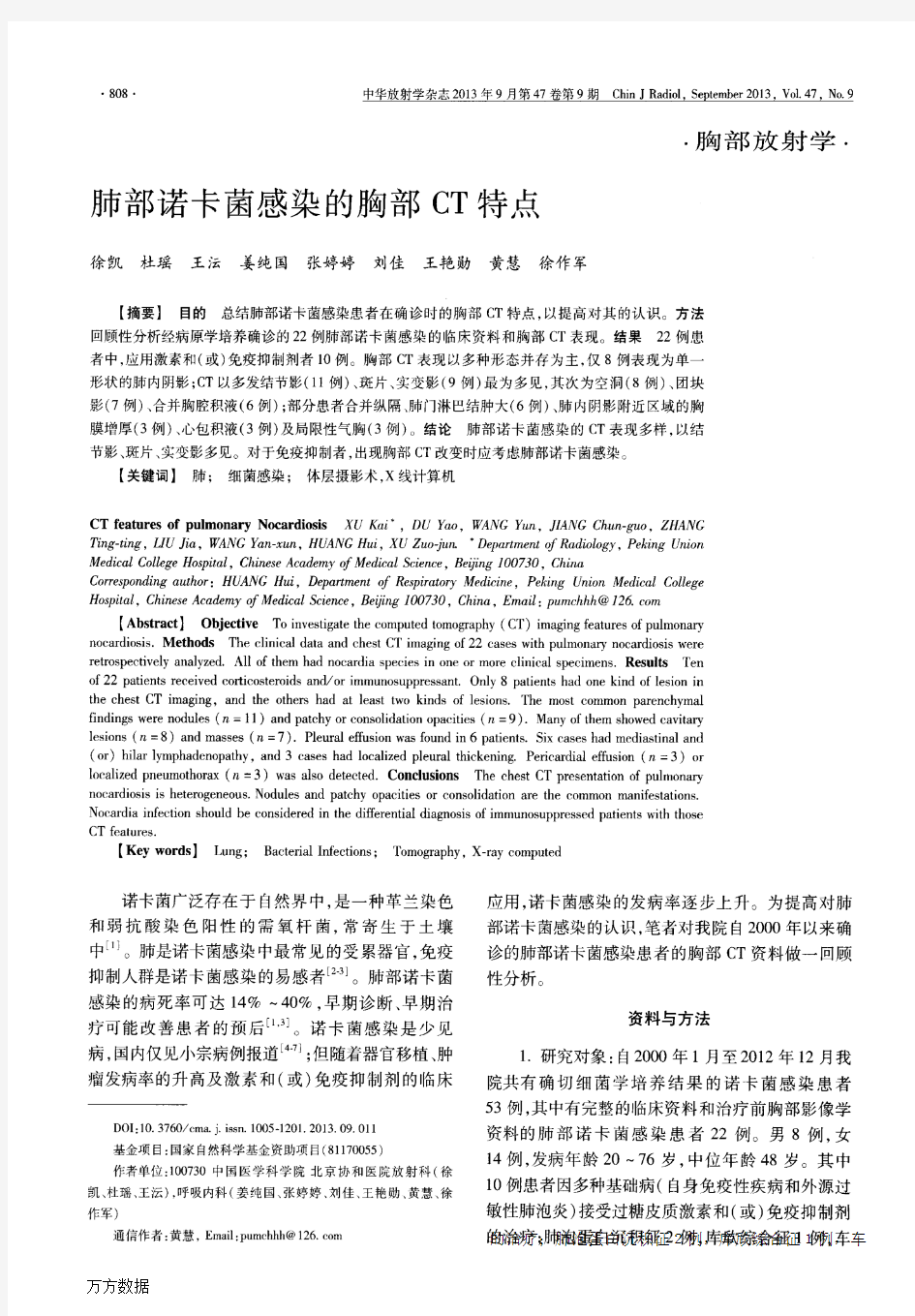
肺部诺卡菌感染的胸部CT特点
徐凯;杜瑶;王沄;姜纯国;张婷婷;刘佳;王艳勋;黄慧;徐作军
【期刊名称】《中华放射学杂志》
【年(卷),期】2013(047)009
【摘要】目的总结肺部诺卡菌感染患者在确诊时的胸部CT特点,以提高对其的认识.方法回顾性分析经病原学培养确诊的22例肺部诺卡菌感染的临床资料和胸部CT表现.结果22例患者中,应用激素和(或)免疫抑制剂者10例.胸部CT表现以多种形态并存为主,仅8例表现为单一形状的肺内阴影;CT以多发结节影(11例)、斑片、实变影(9例)最为多见,其次为空洞(8例)、团块影(7例)、合并胸腔积液(6例);部分患者合并纵隔、肺门淋巴结肿大(6例)、肺内阴影附近区域的胸膜增厚(3例)、心包积液(3例)及局限性气胸(3例).结论肺部诺卡菌感染的CT 表现多样,以结节影、斑片、实变影多见.对于免疫抑制者,出现胸部CT改变时应考虑肺部诺卡菌感染.%Objective To investigate the computed tomography (CT) imaging features of pulmonary nocardiosis.Methods The clinical data and chest CT imaging of 22 cases with pulmonary nocardiosis were retrospectively analyzed.All of them had nocardia species in one or more clinical specimens.Results Ten of 22 patients received corticosteroids and/or immunosuppressant.Only 8 patients had one kind of lesion in the chest CT imaging,and the others had at least two kinds of lesions.The most common parenchymal findings were nodules (n =11) and patchy or consolidation opacities (n =9).Many of them showed cavitary lesions (n =8) and masses (n =7).Pleural effusion